DVT Ultrasound Scan vs. Traditional Methods: Which is More Effective?
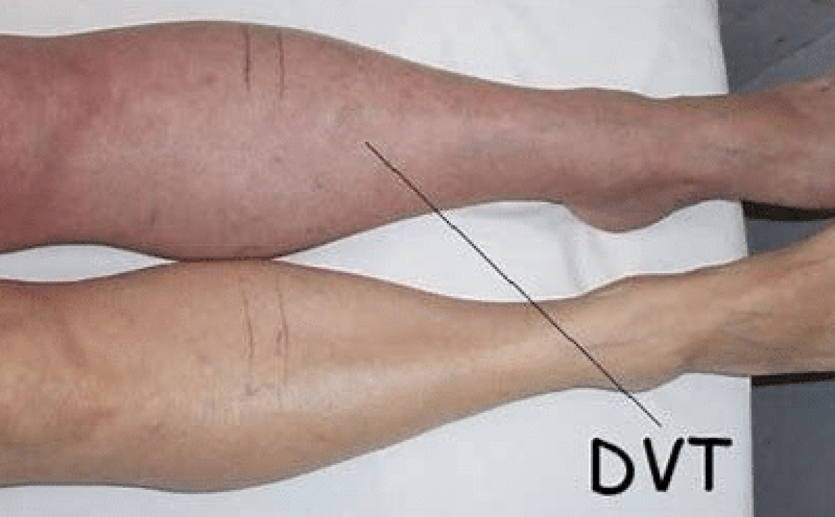
Deep Vein Thrombosis (DVT) is a serious medical condition that occurs when a blood clot forms in a deep vein, most commonly in the legs. If left undetected and untreated, the clot can potentially travel to the lungs, causing a life-threatening condition called pulmonary embolism. To diagnose DVT, healthcare professionals employ various methods, with two predominant ones being ultrasound scans and traditional diagnostic techniques. In this article, we will explore the effectiveness of DVT ultrasound scans compared to the traditional methods, analyzing their respective benefits and limitations to determine which approach is more superior.
Understanding DVT Ultrasound Scans:
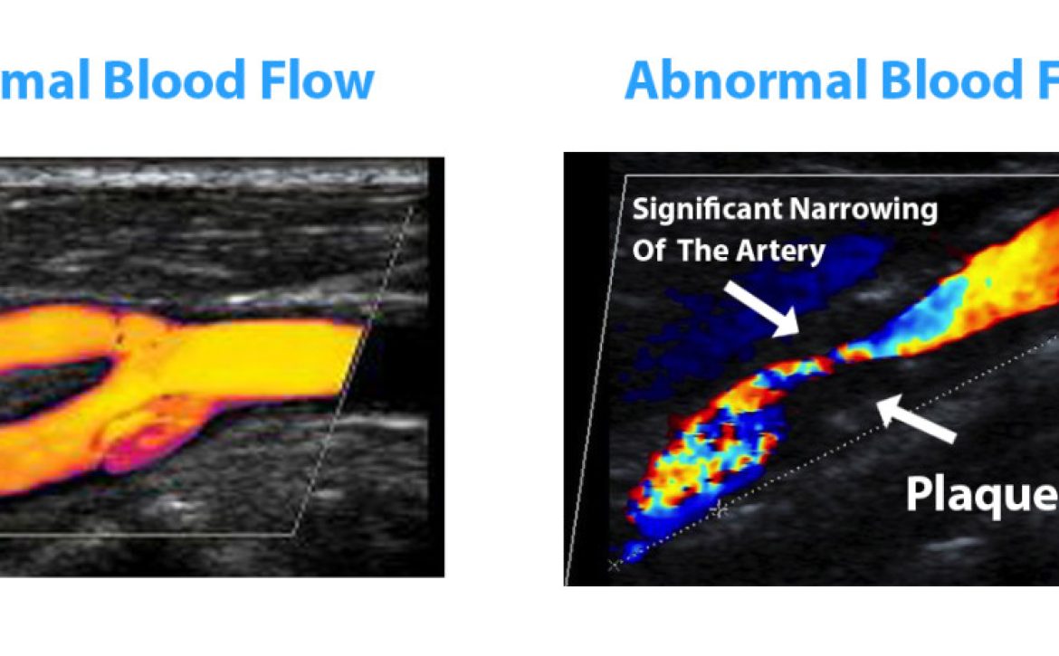
DVT ultrasound scans are non-invasive procedures that utilize sound waves to create images of the veins in the body. During this process, a technician applies a gel to the skin and uses a small handheld device called a transducer to emit sound waves into the body, which bounce back as echoes, creating images of the veins on a monitor. This painless and radiation-free technique is widely considered as the gold standard for diagnosing DVT due to its accuracy and safety.
Advantages of DVT Ultrasound Scans:
-
Accuracy: Studies have shown that DVT ultrasound scans have a high sensitivity and specificity in detecting blood clots. They can identify the presence, location, and size of clots with great precision, allowing healthcare professionals to make informed decisions regarding treatment options.
-
Non-Invasive: Unlike traditional methods such as venography, which involves injecting dye into the veins, ultrasound scans do not require any invasive procedures. This means that patients experience minimal discomfort and have a reduced risk of complications.
-
Real-Time Assessment: DVT ultrasound scans provide real-time imaging, allowing healthcare professionals to visualize the blood flow and assess the condition immediately. This enables prompt treatment initiation, preventing potential complications.
-
Cost-Effective: Compared to traditional methods, DVT ultrasound scans are generally more cost-effective. They do not require hospital admission and can be performed on an outpatient basis, reducing overall medical expenses for both the patients and healthcare systems.
Traditional Methods for DVT Diagnosis:
-
Venography: Venography involves injecting a contrast dye into the veins and taking X-ray images to visualize blood flow. While it provides detailed information on the location and extent of DVT, it is an invasive and potentially uncomfortable procedure.
-
D-dimer Blood Test: A D-dimer blood test measures the levels of a protein fragment released when a blood clot dissolves. While this test can help rule out DVT, it is not specific enough for conclusive diagnosis and often requires additional imaging tests.
Comparing DVT Ultrasound Scans with Traditional Methods:
DVT ultrasound scans have emerged as the preferred diagnostic method for identifying DVT, primarily due to their non-invasiveness, accuracy, and real-time assessment capabilities. They offer numerous advantages over traditional methods, including a reduced risk of complications, cost-effectiveness, and wider availability. However, it is important to acknowledge the limitations of ultrasound scans, especially regarding operator-dependent interpretations and challenges in visualizing clots in certain deep veins.
Conclusion:
In conclusion, DVT ultrasound scans have revolutionized the diagnosis of deep vein thrombosis by providing an accurate, non-invasive, and real-time assessment of blood clots. While traditional methods still have a role in specific cases, the advantages offered by ultrasound scans make them the preferred choice for most healthcare professionals. If you suspect DVT, consult your healthcare provider, who can guide you in determining the most appropriate diagnostic method based on your individual circumstances.


