27 Welbeck Street, London W1G 8EN
Tel: 020 7101 3377
September 2, 2023 by London Private Ultrasound 0 Comments

The Advantages of Elbow Ultrasound Scans Revealed

Introduction: Elbow injuries and conditions can significantly impact an individual’s mobility and overall quality of life. In such cases, accurate diagnosis becomes crucial for effective treatment. While there are various diagnostic imaging techniques available, including X-rays and MRI scans, elbow ultrasound scans have emerged as a reliable and advantageous option. In this article, we will delve into the numerous benefits of elbow ultrasound scans and shed light on why they are considered a valuable diagnostic tool in the medical field.

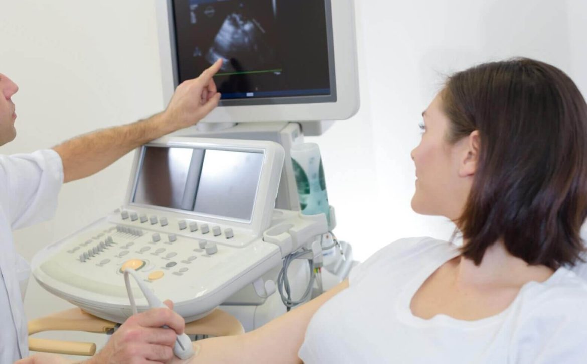
What is Elbow Ultrasound? Elbow ultrasound, also known as sonography or ultrasonography, employs high-frequency sound waves to create detailed images of the internal structures of the elbow joint. By using a handheld transducer and a gel that helps transmit sound waves, medical professionals can visualize the bones, muscles, tendons, and ligaments in real-time.

Benefits of Elbow Ultrasound Scans:

  1. Non-invasive and Safe: Unlike other imaging techniques, such as X-rays or CT scans, elbow ultrasound scans do not expose patients to harmful ionizing radiation. This makes them a safer option, especially for individuals who may require repeated imaging or those more susceptible to radiation-related risks, such as children and pregnant women.

  2. Real-time Visualization: One of the significant advantages of elbow ultrasound scans is the ability to visualize the elbow joint in real-time. Medical professionals can observe the structures while they are in motion, providing valuable insights into any abnormalities or dysfunctional movements. This dynamic visualization aids in diagnosing various conditions accurately and monitoring treatment progress.

  3. High Precision and Detail: With advanced technology, elbow ultrasound scans have become increasingly accurate and detailed. They provide exceptional resolution, allowing clinicians to identify even subtle abnormalities, such as tears in tendons, inflammation, or fluid accumulation. The detailed images obtained contribute to precise diagnosis and guide appropriate treatment plans.

  4. Cost-effective: Compared to other imaging modalities like MRI or CT scans, elbow ultrasound scans are relatively more affordable. Additionally, they can be performed in clinics or medical facilities, eliminating the need for hospital visits or extended stays. The cost-effectiveness of elbow ultrasounds makes them accessible to a wider range of patients, streamlining the diagnostic process.

  5. No Special Preparations Needed: Unlike some imaging techniques, elbow ultrasound scans require minimal preparations. Patients do not need to fast or undergo any special dietary restrictions before the procedure. This convenience adds to the overall patient experience and eliminates any unnecessary anxiety or inconvenience associated with complex preparations.

  6. Versatile and Comprehensive: Elbow ultrasound scans can provide a comprehensive assessment of various conditions, including but not limited to:

  • Tennis or Golfer’s elbow
  • Elbow bursitis
  • Ligament and tendon tears
  • Arthritis
  • Tumors or cysts within the elbow joint
  • Inflammation or fluid accumulation

By obtaining a clear and holistic view of the affected area, medical professionals can devise appropriate treatment plans tailored to the specific condition.

Conclusion: Elbow ultrasound scans have revolutionized the field of diagnostic imaging by offering several advantages over traditional methods. The non-invasive nature, real-time visualization, high precision, cost-effectiveness, and versatility make them a valuable tool in diagnosing and monitoring elbow conditions. With their ability to provide detailed and accurate imaging, elbow ultrasound scans have become a crucial component in the armamentarium of medical professionals, optimizing patient care and facilitating effective treatment plans. If you are experiencing elbow pain or suspect an injury, consult with a healthcare provider who can determine if an ultrasound scan is appropriate for your case.

September 1, 2023 by London Private Ultrasound 0 Comments

Everything You Need to Know about Lumps and Bumps Ultrasound Scan

Introduction:

Lumps and bumps can often be a cause for concern, causing anxiety and worry. In order to properly diagnose and address these issues, medical professionals often recommend an ultrasound scan. This non-invasive procedure has proven to be an effective way to identify and assess lumps and bumps, aiding in early detection and accurate diagnosis. In this article, we will delve into the world of lumps and bumps ultrasound scan, exploring its benefits, procedure, and what you can expect during your appointment.

Understanding Lumps and Bumps Ultrasound Scan:

A lumps and bumps ultrasound scan is a medical imaging technique that uses sound waves to create detailed pictures of the inside of your body. Unlike X-rays or CT scans, ultrasound scans do not involve the use of radiation, making them a safer option, especially for pregnant women or children. This procedure is commonly used to examine various areas of the body, including the breasts, neck, abdomen, and limbs.

Procedure:

Before the scan, your sonographer will explain the process to you and answer any questions you may have. You will be asked to lie down on an examination table, and a gel will be applied to the area being scanned. The sonographer will then use a small handheld device called a transducer to pass over the gel-covered area. This device emits high-frequency sound waves, which bounce back and create images on a monitor. The entire process is painless and typically takes around 15 to 30 minutes, depending on the area being examined.

Benefits of Lumps and Bumps Ultrasound Scan:

  1. Non-invasive: Unlike other imaging techniques, ultrasound scans are non-invasive, meaning they do not require any incisions or injections. This makes them a preferred choice for patients who prefer a less invasive approach.

  2. Safe and radiation-free: As mentioned earlier, ultrasound scans do not involve the use of radiation, making them safe for individuals of all ages, including pregnant women and children.

  3. Accurate and detailed imaging: Ultrasound scans provide real-time images of the area being examined, allowing the sonographer to view the internal structures and identify any abnormalities accurately. This helps in the early detection of potential health issues.

  4. Versatility: Lumps and bumps ultrasound scans can be used to examine various parts of the body, making them a versatile tool in the field of diagnostic medicine.

Determining Lumps and Bumps Differences with a Scan

  1. Breast examination: Ultrasound scans play a crucial role in the evaluation of breast lumps. It helps in distinguishing between lump and bump differences of solid masses and fluid-filled cysts, aiding in the diagnosis of breast cancer.

  2. Thyroid evaluation: Ultrasound scans are often used to assess the thyroid gland for any abnormalities, again this Thyroid ultrasound allows us to identify the lump and bump difference that occurs in nodules or cysts, for example.

  3. Abdominal evaluation: To investigate abdominal pain or detect abnormalities in the liver, gallbladder, pancreas, or kidneys, ultrasound scans are commonly recommended.

  4. Musculoskeletal examination: Ultrasound scans can be used to evaluate various musculoskeletal conditions, such as tendonitis, muscle tears, and joint inflammation. The causes will show up as a lump and bump difference in the mass type, density and manoeuvrability.

Monitor different types of Lumps and Bumps with ultrasound

Obtaining a lumps and bumps ultrasound scan with London Private Ultrasound is a valuable tool in the diagnosis and monitoring of various health conditions. Its non-invasive nature, safety, and versatility make it an ideal choice for patients seeking a detailed and accurate evaluation of lumps and bumps.

If you notice any abnormalities or have concerns about lumps or bumps on your body, it is essential to take action and you are welcome to opt for one of the many ultrasound health scans and lump and bump scans we provide. Alternatively, you may prefer to book a consultation with us first – this can be via an online channel or in our specialist ultrasound clinic where we conduct ultrasounds throughout each and every day with specialised clinics for each type of medical issue. If you are wanting to find out more about what syptoms and signs may indicate cancer we can offer you the support and consultations alongside these ultrasound services.

September 1, 2023 by London Private Ultrasound 0 Comments

Why an Elbow Ultrasound Scan is Vital for Diagnosis

In the world of medical diagnostics, advancements in technology have played a significant role in improving accuracy and efficiency. One such technology that has revolutionized the field is the elbow ultrasound scan. By harnessing the power of sound waves, this non-invasive procedure has become a vital tool for diagnosing various conditions affecting the elbow joint.

So, why is an elbow ultrasound scan so important in the diagnosis process? Let’s explore its benefits and the role it plays in identifying and treating elbow-related conditions.

First and foremost, an elbow ultrasound scan provides healthcare professionals with a detailed image of the elbow joint’s internal structures. By using high-frequency sound waves, the scan captures real-time images, allowing for a comprehensive examination of the joint’s anatomy. This imaging technique helps in detecting abnormalities, fluid accumulation, growths, or any other structural damages that may be affecting the elbow joint.

One of the primary advantages of an ultrasound scan is its ability to provide real-time imaging. Unlike other imaging techniques, such as X-rays or magnetic resonance imaging (MRI), which may require additional time for development or interpretation, ultrasound produces immediate results. The real-time images allow healthcare professionals to visualize the elbow joint in motion, evaluating its functionality and detecting any abnormalities that may only occur during specific movements or positions.

Furthermore, an elbow ultrasound scan is a non-invasive procedure that eliminates the need for surgical intervention. Traditionally, obtaining an accurate diagnosis for elbow conditions required invasive procedures or exploratory surgeries, which are not only costly but also pose certain risks and complications. With the advent of ultrasound technology, healthcare professionals can now perform a thorough assessment of the elbow joint without subjecting the patient to unnecessary invasive procedures.

Another significant advantage of an elbow ultrasound scan is its cost-effectiveness. Compared to other imaging techniques, such as MRI or computed tomography (CT) scans, ultrasound scans are relatively more affordable. This affordability makes it accessible to a broader range of patients, ensuring that everyone has the opportunity to receive a timely and accurate diagnosis for their elbow-related conditions.

Now, let’s take a closer look at some specific elbow conditions that greatly benefit from an ultrasound scan.

Tennis elbow, medically known as lateral epicondylitis, is a common overuse injury affecting the tendons in the elbow region. An ultrasound scan helps in visualizing the inflamed or damaged tendons, allowing for proper identification and targeted treatment. It also assists in differentiating tennis elbow from other similar conditions, such as golfer’s elbow or radial tunnel syndrome.

Golfer’s elbow, or medial epicondylitis, is another condition that can be effectively diagnosed using an elbow ultrasound scan. By capturing real-time images of the affected area, the scan helps in assessing the extent of the injury, identifying any tendon abnormalities, and guiding appropriate treatment options.

In addition to diagnosing tendon-related conditions, an elbow ultrasound scan is also vital in detecting joint-related issues. Conditions such as arthritis, bursitis, or synovitis can be accurately diagnosed by evaluating the joint space, identifying any erosions or fluid accumulation, and determining the overall health and functionality of the joint.

In conclusion, an elbow ultrasound scan has revolutionized the diagnosis and treatment of various elbow-related conditions. This non-invasive and cost-effective imaging technique provides healthcare professionals with detailed and real-time images of the elbow joint, allowing for accurate identification of structural damages, abnormalities, or fluid accumulation. It plays a crucial role in diagnosing conditions such as tennis elbow, golfer’s elbow, arthritis, bursitis, or synovitis. By incorporating an elbow ultrasound scan into the diagnostic process, healthcare professionals can offer more targeted and effective treatment options to their patients.