27 Welbeck Street, London W1G 8EN
Tel: 020 7101 3377
September 21, 2023 by London Private Ultrasound 0 Comments

Elevate Your Health: Lumps and Bumps Ultrasound Scan

In this modern age, staying proactive about our health has become more essential than ever before. One of the most crucial steps in maintaining a healthy lifestyle involves regular health checkups and screenings. When it comes to detecting and diagnosing lumps and bumps in our bodies, an ultrasound scan has proven to be a valuable tool. In this article, we will explore the significance of a lumps and bumps ultrasound scan, its benefits, and its role in elevating our overall health.

Understanding the Lumps and Bumps Ultrasound Scan: 

A lumps and bumps ultrasound scan is a non-invasive imaging technique that utilizes sound waves to create detailed images of the body’s internal structures. By bouncing sound waves off the body’s tissues, this safe and painless procedure allows healthcare professionals to identify any abnormal growths or masses. Unlike other diagnostic tools, ultrasound scans do not involve radiation, making them an ideal choice for frequent monitoring.

Detecting and Diagnosing Lumps and Bumps: 

Early detection is the key to successful treatment of any abnormal growths in the body. With a lumps and bumps ultrasound scan, medical professionals can thoroughly examine and assess these manifestations without the need for invasive procedures. Whether it’s a lump in the breast, neck, abdomen, or any other part of the body, ultrasound scans provide detailed insights into the nature of the growth, aiding in accurate diagnosis.

Why Choose a Lumps and Bumps Ultrasound Scan?

  1. Safety First: With no exposure to radiation, ultrasound scans are a safe option for frequent checkups and monitoring.
  2. Non-Invasive and Pain-Free: Unlike surgical procedures, ultrasound scans are painless and do not require any incisions or recovery time.
  3. Detailed Imaging: The high-resolution images produced by ultrasound scans provide valuable information about the lumps and their characteristics.
  4. Versatile Application: Ultrasound scans can be used to examine various body regions, including breasts, thyroid, liver, and more, making them a versatile diagnostic tool.
  5. Cost-Effective: Compared to other imaging techniques, ultrasound scans are relatively affordable, making them accessible to a wider population.

The Role of Ultrasound Scans in Health Awareness: 

Regular screenings with a lumps and bumps ultrasound scan play a crucial role in raising health awareness. By detecting abnormalities at an early stage, individuals have a higher chance of receiving timely medical intervention, improving their overall health outcomes. Additionally, ultrasound scans provide an opportunity for healthcare professionals to educate patients about self-examination methods, empowering them to take charge of their health.

Conclusion: In conclusion, a lumps and bumps ultrasound scan is an invaluable tool in the journey towards maintaining optimal health. By providing detailed imaging, safe and painless procedures, and the ability to detect abnormalities at their earliest stages, ultrasound scans allow individuals to take proactive steps towards their well-being. Don’t wait for something uncomfortable to become a bigger concern. Prioritize your health and consider scheduling a lumps and bumps ultrasound scan as part of your ongoing wellness routine!

September 20, 2023 by London Private Ultrasound 0 Comments

How Elbow Ultrasound Scans Aid in Treatment Decisions

In the realm of diagnostic imaging, elbow ultrasound scans have emerged as a valuable tool for clinicians in making informed and accurate treatment decisions. This non-invasive technique offers significant advantages in evaluating various elbow conditions, providing crucial insights that aid in optimizing patient care. In this article, we will delve into the world of elbow ultrasound scans, exploring their benefits and how they play a vital role in treatment decisions.

Understanding Elbow Ultrasound Scans: 

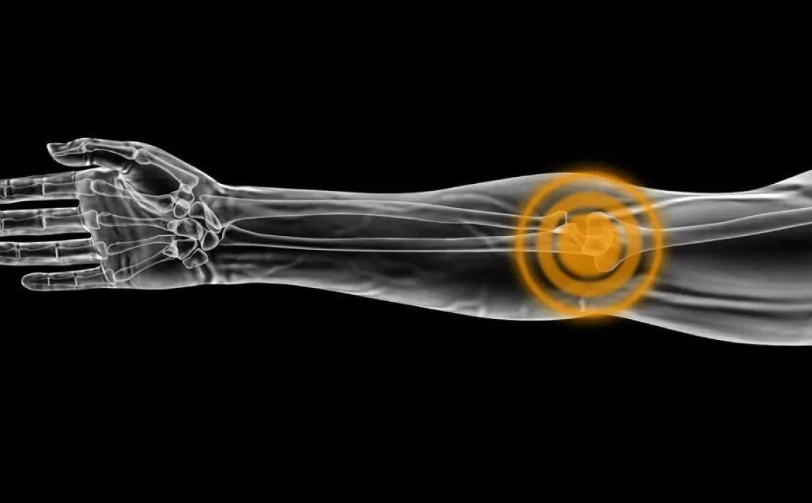
Elbow ultrasound scans employ high-frequency sound waves to create detailed images of the structures within the elbow joint. This imaging technique allows healthcare professionals to visualize and assess the tendons, ligaments, muscles, and other soft tissues in the elbow region. By capturing real-time images, ultrasound scans provide dynamic information about the structure and function of the elbow, facilitating accurate diagnosis and treatment planning.

Benefits of Elbow Ultrasound Scans:

  1. Accurate Diagnosis: Elbow ultrasound scans enable precise diagnosis by providing clear and detailed images of the affected area. This allows healthcare providers to identify injuries, abnormalities, and underlying conditions more effectively, leading to appropriate treatment recommendations.

  2. Non-Invasive and Painless: Unlike other imaging modalities, such as MRI or CT scans, elbow ultrasound is non-invasive and does not involve exposure to radiation. It is a painless procedure that can be performed conveniently in an outpatient setting, ensuring patient comfort and safety.

  3. Real-Time Visualization: One of the most significant advantages of elbow ultrasound scans is the real-time visualization it offers. Physicians can observe the movement of tendons, ligaments, and muscles during various elbow movements, improving the accuracy of the diagnosis and providing valuable insights into the condition.

  4. Versatility: Elbow ultrasound scans are versatile and can be utilized for a wide range of elbow conditions. From diagnosing tendinitis, bursitis, and arthritis to evaluating injuries such as fractures, sprains, and subluxations, this imaging technique provides comprehensive information for treatment planning.

  5. Cost-Effective: Compared to other imaging modalities, elbow ultrasound scans are relatively cost-effective. This affordability allows for wider accessibility to diagnostic services, ensuring that patients receive timely and accurate evaluations of their elbow conditions.

Role in Treatment Decisions: 

Elbow ultrasound scans play a crucial role in treatment decisions by providing valuable insights that guide healthcare providers in determining the most effective course of action. Here’s how these scans aid in treatment planning:

  1. Accurate Localization: Elbow ultrasound scans help localize the precise site of the injury or condition, enabling targeted treatments such as injections or physical therapy. By identifying the exact location of the problem, clinicians can administer therapies directly to the affected area, optimizing treatment outcomes.

  2. Evaluation of Tissue Healing: Monitoring the progress of tissue healing is vital for assessing treatment effectiveness. Elbow ultrasound scans can be used to observe the healing process over time, allowing clinicians to modify treatment plans or interventions as needed.

  3. Guiding Aspiration and Injections: Elbow ultrasound scans assist in guiding the aspiration of fluid or the administration of injections to the affected area. This ensures precision and reduces the risk of complications, improving patient comfort and outcomes.

Conclusion: Elbow ultrasound scans have become an invaluable tool in the field of diagnostic imaging, offering numerous benefits for both healthcare providers and patients. This non-invasive technique aids in accurate diagnosis, treatment planning, and monitoring the healing process. By utilizing elbow ultrasound scans, clinicians can optimize treatment decisions, ensuring the best possible outcomes for their patients. If you’re experiencing elbow pain or suspect an elbow condition, consult with your healthcare provider about the potential benefits of an elbow ultrasound scan.

September 18, 2023 by London Private Ultrasound 0 Comments

Discovering the Power of Lumps and Bumps Ultrasound Scan

 Ultrasound scans have become an essential tool in modern medical diagnostics, enabling healthcare professionals to detect and monitor a wide range of conditions. One such application is the detection and evaluation of lumps and bumps. In this article, we will explore the power of lumps and bumps ultrasound scans, their benefits, and their role in diagnosing various medical conditions. Let’s dive in!

Understanding Ultrasound Scans for Lumps and Bumps: 

Ultrasound scans use high-frequency sound waves to create detailed images of the body’s internal structures, without the need for invasive procedures. When it comes to lumps and bumps, ultrasound scans provide valuable information about the composition, size, and location of abnormalities, aiding in accurate diagnosis and treatment planning.

  1. The Importance of Early Detection: Early detection of lumps and bumps is crucial for effective medical intervention. Ultrasound scans allow healthcare professionals to identify abnormalities that may not be visible or detectable through other means. By spotting these changes at an early stage, healthcare providers can initiate appropriate treatment protocols, potentially saving lives.

  2. Evaluating Soft Tissue Abnormalities: Soft tissue abnormalities, such as cysts, tumors, or benign growths, can often present as lumps or bumps. Ultrasound scans offer a non-invasive way to evaluate these abnormalities, providing detailed images of the affected area. By examining the shape, size, and characteristics of the lump or bump, medical professionals can differentiate between benign and malignant growths and determine the best course of action.

  3. Guiding Biopsies and Fine Needle Aspiration: In cases where a lump or bump requires further investigation, ultrasound scans can guide biopsy procedures or fine needle aspiration. By using real-time imaging, healthcare providers can precisely target the abnormality, ensuring accurate sample collection. This minimally invasive approach reduces the risks associated with more invasive procedures while providing essential diagnostic information.

  4. Monitoring Treatment Progress: Ultrasound scans are not only valuable for initial diagnosis but also for monitoring the progress of treatment. After interventions like surgeries, chemotherapy, or radiation therapy, regular ultrasound scans assist healthcare professionals in assessing the effectiveness of the treatment, identifying any recurrence or new developments, and adjusting the treatment plan accordingly.

  5. Ultrasound vs. Other Imaging Techniques: Compared to other imaging techniques like X-rays or computed tomography (CT) scans, ultrasound scans offer several advantages when evaluating lumps and bumps. Ultrasound scans do not use ionizing radiation, making them safer for both patients and medical professionals. Additionally, ultrasound scans provide real-time imaging, allowing healthcare providers to assess dynamic changes within the lump or bump immediately.

Conclusion: Lumps and bumps ultrasound scans have revolutionized the way medical professionals approach the detection and evaluation of various abnormalities. These non-invasive procedures offer a wealth of information, aiding in early detection, proper diagnosis, and treatment monitoring. If you suspect any lumps or bumps, consult your healthcare provider to determine whether an ultrasound scan is warranted. Remember, early detection can make a significant difference in your health outcomes.

September 16, 2023 by London Private Ultrasound 0 Comments

Elbow Ultrasound vs. Other Diagnostic Methods: Making the Right Choice

When it comes to diagnosing elbow injuries and conditions, medical professionals have a range of diagnostic methods to choose from. One method that has gained popularity in recent years is elbow ultrasound. In this article, we will explore the benefits of elbow ultrasound compared to other diagnostic methods, and discuss why it is the right choice for accurate and efficient diagnosis.

Understanding Elbow Ultrasound:

Elbow ultrasound utilizes sound waves to create images of the structures within the elbow joint. This non-invasive procedure allows doctors to assess the tendons, ligaments, muscles, and other soft tissues in real-time. The use of ultrasound in diagnosing elbow injuries has several advantages over traditional methods such as X-rays, CT scans, and MRI scans.

Advantages of Elbow Ultrasound:

  1. Non-invasive and Safe: Elbow ultrasound does not involve ionizing radiation like X-rays and CT scans, making it a safer option for patients, especially for those who require multiple imaging sessions.

  2. Real-time Imaging: Unlike MRI scans, which require the patient to remain still for an extended period, elbow ultrasound provides real-time imaging. This means that doctors can assess the elbow joint while it is in motion, providing valuable insights into dynamic conditions such as ligament tears or joint instability.

  3. Cost-effective: Elbow ultrasound is generally more cost-effective than other imaging techniques, making it a preferred choice for both patients and healthcare providers. This affordability allows for repeated imaging sessions when necessary, leading to better monitoring of treatment progress.

  4. Availability and Accessibility: Elbow ultrasound machines are widely available in medical facilities, and the procedure can be performed by trained technicians. This accessibility ensures that patients can receive timely and accurate diagnoses without long wait times or travel to specialized imaging centers.

Comparing Elbow Ultrasound to Other Diagnostic Methods:

While elbow ultrasound has numerous advantages, it is important to acknowledge the limitations and situations where alternative diagnostic methods may be more appropriate. Let’s compare elbow ultrasound to some commonly used imaging techniques:

  1. X-rays: X-rays are useful for evaluating bone fractures and bone-related conditions. However, they are less effective in assessing soft tissue injuries, such as tendon or ligament tears, making elbow ultrasound a superior choice for these cases.

  2. CT Scans: CT scans provide detailed cross-sectional images and are valuable for assessing complex fractures or bone abnormalities. However, they involve radiation exposure and are not as suitable for soft tissue evaluation as elbow ultrasound.

  3. MRI Scans: MRI scans offer high-resolution images of soft tissues, making them a preferred choice for detecting subtle injuries or evaluating complex conditions like ligament tears. However, they can be expensive and time-consuming compared to elbow ultrasound.

Conclusion:

In conclusion, elbow ultrasound is a safe, cost-effective, and readily available diagnostic method that offers real-time imaging and accurate assessments of soft tissue injuries in the elbow joint. While other diagnostic methods have their merits in specific scenarios, elbow ultrasound stands out due to its non-invasiveness, affordability, accessibility, and ability to capture dynamic conditions. If you or someone you know is experiencing elbow pain or injury, consult a medical professional who can guide you in making the right diagnostic choice.

September 11, 2023 by London Private Ultrasound 0 Comments

Step-by-Step Guide: What to Expect during an Elbow Ultrasound Scan

In the world of medical imaging, ultrasound scans have become indispensable tools for diagnosing and monitoring various conditions. Although commonly known for their use in prenatal examinations, ultrasound scans are also employed to assess musculoskeletal problems, including those affecting the elbow joint. In this comprehensive guide, we will take you through the step-by-step process of an elbow ultrasound scan, shedding light on its importance, procedure, and what to expect during the examination.

I. Understanding Elbow Ultrasound Scans:

Before delving into the specifics, let’s establish a basic understanding of elbow ultrasound scans and their significance. Elbow ultrasound scans utilize sound waves to create real-time images of the soft tissues, bones, and cartilage within and around the elbow joint. They are non-invasive, safe, and provide valuable information to physicians, helping them diagnose and treat various conditions such as tennis elbow, golfer’s elbow, bursitis, or ligament injuries.

II. Preparing for the Elbow Ultrasound Scan:

  1. Research and Choose a Reputable Imaging Facility:

    • Research the best imaging facilities in your area that offer elbow ultrasound scans.
    • Look for facilities with experienced radiologists and modern equipment.
  2. Schedule an Appointment:

    • Call the chosen facility and schedule an appointment for your elbow ultrasound scan.
    • Inquire about any specific instructions or preparations needed for the examination.
  3. Dress Comfortably:

    • Wear loose-fitting clothing that allows easy access to your elbow.
    • Avoid wearing jewelry or accessories around the examination area.

III. The Procedure:

  1. Arrival and Check-in:

    • Arrive at the imaging facility a few minutes before your scheduled appointment.
    • Complete any necessary paperwork and check-in with the receptionist.
  2. Room Preparation:

    • A technologist or radiologist will guide you to the examination room.
    • They will explain the procedure and answer any questions you may have.
  3. Positioning:

    • You’ll be asked to lie on your back or sit comfortably, depending on the examination type.
    • The radiologist may provide pillows or position your arm to ensure optimal imaging.
  4. Gel Application:

    • The radiologist applies a clear, water-based gel to your elbow area.
    • The gel helps transmit sound waves and enhances image quality.
  5. Transducer Placement and Scanning:

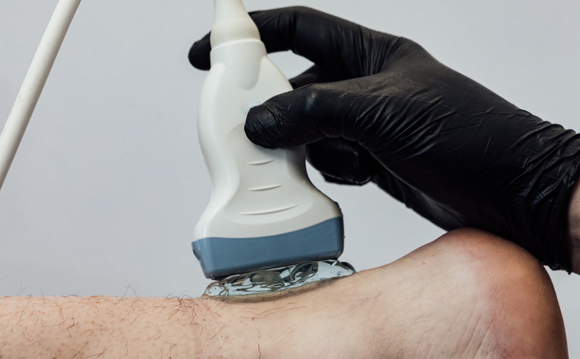
    • The radiologist gently presses a handheld transducer against your elbow.
    • The transducer emits sound waves and captures the returning echoes, forming images on a screen.
  6. Image Interpretation:

    • The radiologist analyzes the real-time images, assessing the structures of your elbow.
    • They may ask you to move your arm or flex your muscles to evaluate specific areas.
  7. Duration:

    • The exact time may vary depending on your unique case and the radiologist’s evaluation.

IV. Conclusion:

Undergoing an elbow ultrasound scan is a vital step in diagnosing and managing elbow-related conditions. By following this step-by-step guide, you are now equipped with the knowledge of what to expect during the examination. Remember to schedule your appointment at a reputable imaging facility, dress comfortably, and be prepared for the scanning process.

Back psoriasis. Man hands scratching the itch on the back. Concept of health and medicine
September 9, 2023 by London Private Ultrasound 0 Comments

5 Warning Signs You Shouldn’t Ignore: Lumps and Bumps Ultrasound Scan

Lumps and Bumps Diagnosis with Ultrasound

Lumps and bumps on the body can be a cause for concern. While some may be harmless, others might signal a more serious underlying health condition. Ignoring these warning signs can lead to delayed diagnosis of lumps and bumps and the correct course of treatment, potentially impacting your overall well-being. In this article, we will discuss five warning signs that you should never ignore when it comes to lumps and bumps, highlighting the importance of ultrasound scans in detecting and diagnosing potential health issues.

Lump and Bump Symptoms

  1. Unexplained Growth or Enlargement:

One of the first warning signs you shouldn’t ignore is the sudden appearance or significant enlargement of a lump or bump. Whether it’s on your skin, under your armpit, or within your breast tissue, any unexplained growth should be examined by a healthcare professional. An ultrasound scan can provide detailed images, allowing experts to determine the nature of the growth and guide further investigations if necessary.

  1. Changes in Shape or Texture:

Pay close attention to any changes in the shape or texture of lumps and bumps. If a previously smooth surface becomes irregular or starts to feel harder, it could be indicative of a problem. An ultrasound scan can help evaluate the internal structure of the mass, providing valuable insights into its composition and potential abnormalities.

  1. Persistent Pain or Discomfort:

Experiencing persistent pain or discomfort around a lump or bump should never be ignored. While not all painful lumps are cancerous, it’s crucial to have them properly assessed. An ultrasound scan can assist in identifying potential causes of the pain, such as inflammation or tissue damage, and aid in the accurate diagnosis.

  1. Rapid Growth and Unusual Coloration:

If a lump or bump exhibits rapid growth or displays unusual coloration, seeking medical attention is imperative. These signs might indicate an infection, a benign tumor, or even a malignant growth. Ultrasound scans can help differentiate between various types of growths and assist in determining whether further interventions, such as a biopsy, are required.

  1. Changes in Nearby Lymph Nodes:

Lumps and bumps often coincide with changes in nearby lymph nodes. If you notice the nodes becoming swollen, tender, or hard, it could be an indication of an underlying issue. An ultrasound scan can help evaluate the lymph nodes’ size, shape, and texture, providing valuable information to aid in diagnosis and treatment planning. We offer a full body lymph node scan and options for one area and a lymph node scan of two areas

London Private Ultrasound Lump and Bump Appointments

When it comes to lumps and bumps, staying vigilant and recognizing the warning signs can be life-saving. By paying attention to unexplained growth, changes in shape or texture, persistent pain or discomfort, rapid growth with unusual coloration, and changes in nearby lymph nodes, you can take proactive steps towards a timely diagnosis and potential treatment. Remember, an ultrasound scan is an essential tool in identifying and evaluating these warning signs, providing medical professionals with the information needed to make accurate diagnoses and recommendations for further action.

LPU is a specialist ultrasound clinic in Central London, we have a team of experienced practitioners and clinicians able to guide and advise you, provide consultations and after the scan/s and screenings can produce reports and accurate diagnosis. We have additional services such as Blood Testing and specislists in the field of cardiovascular health, cardiology, pregnancy and MSK as well as those who are experienced with cancer, mens and womens health and much more. We aim to provide a suite of services for those needing fast and accurate diagnosis when experiencing worrying signs or symptoms as well as providing health maintenance or seeking the medical information to achieve optimal health or needing to sustain advanced training routines.

September 7, 2023 by London Private Ultrasound 0 Comments

Mastering the Diagnosis: Lumps and Bumps Ultrasound Scan

In today’s fast-paced world, it is essential to have advanced diagnostic tools that aid healthcare professionals in accurate and timely diagnoses and lumps and bumps ultrasound reports play a vital role in that process. One such tool that has revolutionised the field of medical imaging is ultrasound imaging. This non-invasive and safe technique allows physicians to visualize and assess abnormalities beneath the skin’s surface. In this article, we will delve into the details of a lumps and bumps ultrasound scan, its significance in diagnosing various conditions, and how it has become an indispensable tool for healthcare providers.


Understanding the Lumps and Bumps Ultrasound Scan

A lumps and bumps ultrasound scan, also known as a soft tissue ultrasound or superficial ultrasound, is a medical imaging procedure that employs high-frequency sound waves to produce real-time images of the body’s soft tissues. Unlike other imaging techniques, such as X-rays or CT scans, ultrasound uses sound waves instead of radiation, making it exceptionally safe for both patients and healthcare professionals and provides the opportunity to fully examine the tissue for the purpose of generating lumps and bumps ultrasound reports.

The Importance of Ultrasound in Diagnosis

  1. Accurate Identification and Localization: The lumps and bumps ultrasound scan excels in accurately identifying the location and composition of various masses beneath the skin. This enables healthcare professionals to rapidly diagnose conditions such as cysts, lipomas, abscesses, and identify cancerous tumors. With ultrasound, physicians can determine whether a lump is fluid-filled or solid, aiding in appropriate treatment planning.

  2. Real-time Visualization: Real-time imaging capabilities provided by ultrasound allow healthcare providers to visualize dynamic movements within the body. This feature proves particularly useful during ultrasound-guided procedures, such as fine-needle aspiration or biopsies, ensuring precise sampling and reducing potential complications.

  3. Non-invasiveness and Patient Comfort: Unlike invasive procedures like surgical biopsies, lumps and bumps ultrasound scans are non-invasive and cause minimal discomfort. This makes it an ideal choice, especially for sensitive areas, pediatric patients, and individuals who may not be candidates for surgical procedures.

  4. Cost-effectiveness: The lumps and bumps ultrasound scan is relatively cost-effective compared to other imaging modalities, such as magnetic resonance imaging (MRI) or computed tomography (CT). This affordability factor makes it accessible to a broader population, facilitating early diagnosis and improving patient outcomes.

We provide urgent same day clinics and provide a fast-track service to create a lumps and bumps ultrasound report and also provide opportunity to seek GP consultation and guidance so that you fully understand the findings and what they mean.

Usage and Applications

The applications of lumps and bumps ultrasound scans are diverse, ranging from routine examinations to the detection and monitoring of diseases. Let’s explore a few prominent examples:

  1. Musculoskeletal Conditions: Ultrasound is widely used to assess musculoskeletal conditions, including assessing joint effusions, diagnosing sprains or tears in ligaments and tendons, and evaluating soft tissue masses such as ganglion cysts or lipomas.

  2. Breast Examinations: Lumps and bumps ultrasound scans play a crucial role in breast imaging, aiding in the detection, characterization, and follow-up of breast masses. Ultrasound-guided biopsies are also performed to obtain tissue samples, guiding subsequent treatment decisions.

  3. Thyroid Evaluations: Ultrasound imaging effectively visualizes the thyroid gland, facilitating the diagnosis of thyroid nodules, goiters, and assessing their characteristics. It is especially valuable in distinguishing between benign and malignant nodules, leading to appropriate management.

Lumps and bumps ultrasound reports

Mastering the Diagnosis: Lumps and Bumps Ultrasound Scan is an indispensable tool in modern medicine. Its non-invasive nature, accuracy in identifying and localising abnormalities, real-time imaging, and cost-effectiveness make it a preferred choice for healthcare professionals. By giving insights into the world of lumps and bumps ultrasound reporting, the investigations and evaluations we hope to have highlighted its significance in diagnosing various conditions effectively. 

September 5, 2023 by London Private Ultrasound 0 Comments

The Ultimate Guide to Lumps and Bumps Ultrasound Scan

If you’ve ever noticed an unusual lump or bump on your body, it’s only natural to feel concerned. The fear of the unknown can be overwhelming. Fortunately, medical technology has advanced considerably, providing a non-invasive and effective method of diagnosing lumps and bumps—ultrasound scans. In this comprehensive guide, we’ll explore everything you need to know about lumps and bumps ultrasound scans, including their benefits, procedure, and potential diagnoses.

Understanding Lumps and Bumps

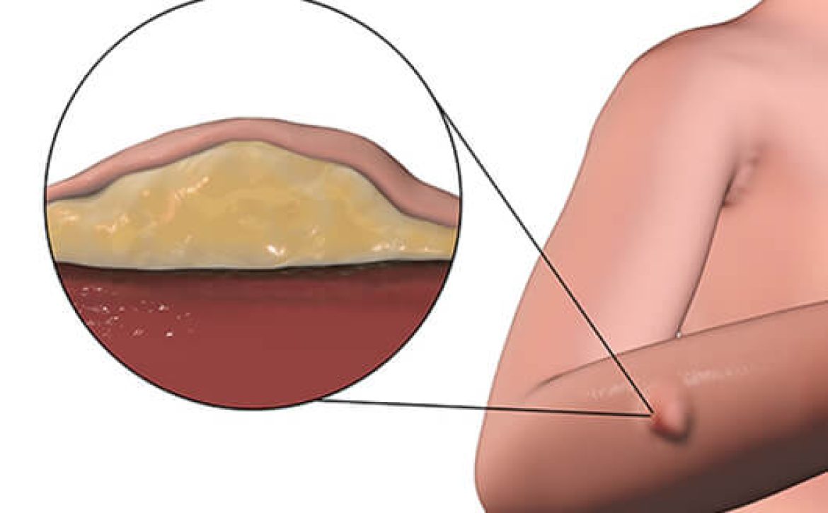
Before diving into ultrasounds, let’s gain a basic understanding of lumps and bumps. These can vary in shape, size, and texture, and may occur on any part of the body. Common causes include cysts, lipomas (benign fatty tumors), swollen lymph nodes, or even cancerous growths. Detecting the nature of the lump is vital for accurate diagnosis and appropriate treatment.

Benefits of Ultrasound Scans

Ultrasound scans have become widely used due to several key advantages. Firstly, they are safe and painless, involving no exposure to ionizing radiation. Secondly, they provide real-time imaging, allowing for immediate evaluation, which is especially valuable during the scan. Lastly, ultrasound scans are accessible and cost-effective, making them the go-to procedure for assessing lumps and bumps.

The Ultrasound Procedure

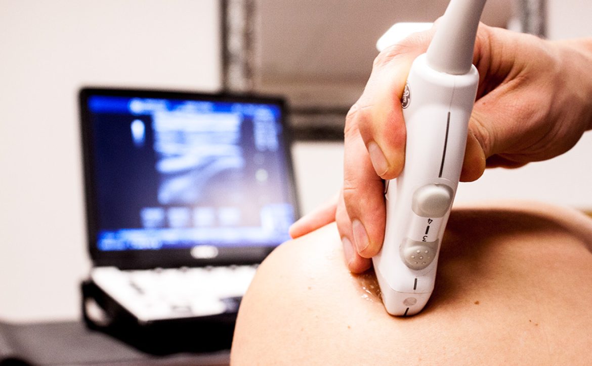
When you arrive for your ultrasound scan, you’ll be guided to a comfortable examination room. A trained sonographer or radiologist will apply a gel to your skin, which helps transmit the sound waves and eliminate air pockets. The specialist will then gently glide a small handheld device called a transducer over the area of concern. This device emits high-frequency sound waves, which bounce off the tissues and create echoes that are captured and transformed into images by the ultrasound machine.

Diagnosing Lumps and Bumps

Thanks to advancements in ultrasound technology, medical professionals can determine the nature of lumps and bumps with remarkable accuracy. Ultrasound scans can identify fluid-filled cysts, distinguish between solid and fatty masses, evaluate the vascularity of tissues, and assist in guiding needle biopsies when further investigation is required. The ability to visualize the internal structures helps in reaching a precise diagnosis.

Common Diagnoses

  1. Cysts: Often filled with fluid, cysts are typically benign and often require no treatment. Ultrasound scans can accurately locate and measure cysts, aiding in proper management.

  2. Lipomas: These benign fatty tumors can be easily identified through the characteristic appearance on ultrasound images. The scan helps confirm the diagnosis, ruling out any malignant potential.

  3. Lymph Nodes: Ultrasound scans play a crucial role in assessing enlarged lymph nodes, aiding in the diagnosis of infections, autoimmune conditions, or even malignancy.

  4. Tumors: Whether benign or malignant, ultrasound scans provide valuable information about the size, location, and characteristics of tumors, helping determine the course of treatment.

Conclusion

In conclusion, if you notice any lumps or bumps on your body, consider getting an ultrasound scan for a detailed evaluation. Not only are ultrasound scans safe and painless, but they also offer real-time imaging and accurate diagnoses. By seeking early intervention and professional medical advice, you can ensure the best possible outcome for your health.

September 5, 2023 by London Private Ultrasound 0 Comments

Diagnosing Elbow related issues with Ultrasound Scans

Elbow injuries can be particularly debilitating, affecting people in various age groups and professions. Thankfully, with advancements in medical technology, elbow ultrasound scans have emerged as a non-invasive and effective diagnostic tool. In this article, we will explore the advantages of elbow ultrasound scans and shed light on how they can help in diagnosing and managing elbow-related conditions.

Understanding Elbow Ultrasound Scans:

Elbow ultrasound scans utilize high-frequency sound waves to create detailed images of the internal structures of the elbow joint. This painless procedure is performed by a skilled sonographer or a qualified radiologist. By producing real-time images, elbow ultrasound scans provide valuable insights into potential abnormalities, assisting in accurate diagnosis and efficient treatment planning.

Advantages of Elbow Ultrasound Scans:

  1. Non-Invasive and Safe: Elbow ultrasound scans are non-invasive, meaning they do not require any surgical incisions. Unlike other imaging techniques such as X-rays or CT scans, ultrasound scans do not use ionizing radiation, making them safe for repeat examinations, even for pregnant women and children.

  2. Real-Time Imaging: One significant advantage of elbow ultrasound scans is the ability to observe the elbow joint in real-time. This feature allows healthcare professionals to evaluate joint mobility, identify abnormalities during movement, and assess the response to specific maneuvers or treatments.

  3. Detailed Visualization: Elbow ultrasound scans provide detailed images of the muscles, tendons, ligaments, and other soft tissues of the elbow joint. This level of visualization helps in identifying various conditions, including tendon tears, bursitis, arthritis, and inflammation, leading to more accurate diagnoses.

  4. Guided Procedures: Elbow ultrasound scans can also be used as a guide during various minimally invasive procedures, such as injections or aspirations. The real-time imaging capability assists physicians in precisely targeting the affected area, ensuring the delivery of medications or fluid removal with increased accuracy.

  5. Cost-Effective: Compared to other imaging modalities like MRI or CT scans, elbow ultrasound scans are generally more cost-effective. They offer a viable option for initial evaluation, enabling healthcare providers to assess the condition before recommending further investigative procedures, potentially saving both time and money for the patient.

Conclusion:

In conclusion, elbow ultrasound scans offer numerous advantages in diagnosing and managing elbow-related conditions. With their non-invasive nature, real-time imaging capabilities, and ability to visualize soft tissue structures, these scans have become an indispensable tool for healthcare professionals. By considering the advantages discussed in this article, individuals experiencing elbow pain or injuries can seek appropriate medical attention, leading to accurate diagnoses and effective treatment plans.

September 3, 2023 by London Private Ultrasound 0 Comments

Unveiling the Hidden Truth: Lumps and Bumps Ultrasound Scan

Introduction:

When it comes to medical diagnostic imaging, ultrasound scans have become an integral tool in the detection and evaluation of various conditions. Among these, the identification and examination of lumps and bumps through ultrasound have proven to be highly effective. In this article, we will delve into the hidden truths of lumps and bumps ultrasound scans, demystifying the process and shedding light on its significance in diagnosing underlying health concerns.

Understanding Lumps and Bumps Ultrasound Scans:

Ultrasound scans, also known as sonograms, utilize high-frequency sound waves to create images of the internal structures of the body. These scans offer a non-invasive and safe approach to visualize lumps, bumps, or masses, providing crucial information about their nature and characteristics.

  1. The Importance of Lumps and Bumps Ultrasound Scans:

Lumps and bumps can evoke fear and uncertainty in individuals, often causing anxiety about potential health issues. Ultrasound scans are particularly valuable in reassuring patients or identifying potential concerns, as they can differentiate between solid masses, fluid-filled cysts, or benign tumors. With early detection being vital in many cases, ultrasound scans offer a proactive approach to diagnosis and subsequent treatment.

  1. Indications and Applications:

Lumps and bumps ultrasound scans are used across various medical specialties, serving as a primary diagnostic tool or guiding further explorations. Some common indications for these scans include:

a. Breast Abnormalities: Ultrasound scans help determine the nature of breast lumps or abnormalities detected during self-examinations or mammograms. This method aids in differentiating between benign cysts and potentially malignant masses.

b. Thyroid Nodules: The evaluation of thyroid nodules is another significant use of ultrasound scans. By assessing the size, shape, and internal characteristics of nodules, doctors can decide whether further interventions, such as biopsies, are necessary.

c. Musculoskeletal Lumps: For individuals with palpable lumps or bumps in their muscles or joints, ultrasound scans can provide valuable information about the structures involved, aiding in accurate diagnoses and treatment planning.

d. Abdominal Masses: Ultrasound scans can detect and characterize lumps or masses in the abdominal region, assisting in differentiating between solid tumors, cysts, or other anomalies.

  1. The Ultrasound Process and Techniques:

During a lumps and bumps ultrasound scan, a skilled sonographer or radiologist utilizes a handheld transducer to send and receive sound waves. The transducer is applied to the skin, coupled with a gel that enhances sound wave transmission. The produced images are then analyzed, interpreted, and communicated to the patient by a medical professional.

a. Real-Time Imaging: One of the significant advantages of ultrasound scans is that they offer real-time imaging. This means that the live images allow for dynamic assessments, ensuring a comprehensive evaluation of lumps and bumps.

b. Doppler Ultrasound: In certain cases, when assessing blood flow patterns within lumps or evaluating vascular abnormalities, a Doppler ultrasound technique may be employed. This technique provides additional insights into the vascularity and blood supply of the detected lesions.

Conclusion:

In conclusion, lumps and bumps ultrasound scans provide an invaluable tool in detecting, characterizing, and evaluating various conditions across different medical specialties. These scans help alleviate patient concerns by providing accurate information about the nature and composition of lumps or bumps. By encouraging early intervention and facilitating timely treatment, ultrasound scans play a crucial role in maintaining and promoting overall health.