27 Welbeck Street, London W1G 8EN
Tel: 020 7101 3377
deep vein thrombosis
September 21, 2023 by London Private Ultrasound 0 Comments

DVT Ultrasound Scan vs. Traditional Methods: Which is More Effective?

Deep Vein Thrombosis (DVT) is a serious medical condition that occurs when a blood clot forms in a deep vein, most commonly in the legs. If left undetected and untreated, the clot can potentially travel to the lungs, causing a life-threatening condition called pulmonary embolism. To diagnose DVT, healthcare professionals employ various methods, with two predominant ones being ultrasound scans and traditional diagnostic techniques. In this article, we will explore the effectiveness of DVT ultrasound scans compared to the traditional methods, analyzing their respective benefits and limitations to determine which approach is more superior.

Understanding DVT Ultrasound Scans:

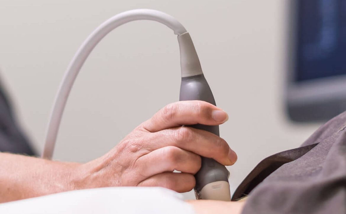
DVT ultrasound scans are non-invasive procedures that utilize sound waves to create images of the veins in the body. During this process, a technician applies a gel to the skin and uses a small handheld device called a transducer to emit sound waves into the body, which bounce back as echoes, creating images of the veins on a monitor. This painless and radiation-free technique is widely considered as the gold standard for diagnosing DVT due to its accuracy and safety.

Advantages of DVT Ultrasound Scans:

  1. Accuracy: Studies have shown that DVT ultrasound scans have a high sensitivity and specificity in detecting blood clots. They can identify the presence, location, and size of clots with great precision, allowing healthcare professionals to make informed decisions regarding treatment options.

  2. Non-Invasive: Unlike traditional methods such as venography, which involves injecting dye into the veins, ultrasound scans do not require any invasive procedures. This means that patients experience minimal discomfort and have a reduced risk of complications.

  3. Real-Time Assessment: DVT ultrasound scans provide real-time imaging, allowing healthcare professionals to visualize the blood flow and assess the condition immediately. This enables prompt treatment initiation, preventing potential complications.

  4. Cost-Effective: Compared to traditional methods, DVT ultrasound scans are generally more cost-effective. They do not require hospital admission and can be performed on an outpatient basis, reducing overall medical expenses for both the patients and healthcare systems.

Traditional Methods for DVT Diagnosis:

  1. Venography: Venography involves injecting a contrast dye into the veins and taking X-ray images to visualize blood flow. While it provides detailed information on the location and extent of DVT, it is an invasive and potentially uncomfortable procedure.

  2. D-dimer Blood Test: A D-dimer blood test measures the levels of a protein fragment released when a blood clot dissolves. While this test can help rule out DVT, it is not specific enough for conclusive diagnosis and often requires additional imaging tests.

Comparing DVT Ultrasound Scans with Traditional Methods:

DVT ultrasound scans have emerged as the preferred diagnostic method for identifying DVT, primarily due to their non-invasiveness, accuracy, and real-time assessment capabilities. They offer numerous advantages over traditional methods, including a reduced risk of complications, cost-effectiveness, and wider availability. However, it is important to acknowledge the limitations of ultrasound scans, especially regarding operator-dependent interpretations and challenges in visualizing clots in certain deep veins.

Conclusion:

In conclusion, DVT ultrasound scans have revolutionized the diagnosis of deep vein thrombosis by providing an accurate, non-invasive, and real-time assessment of blood clots. While traditional methods still have a role in specific cases, the advantages offered by ultrasound scans make them the preferred choice for most healthcare professionals. If you suspect DVT, consult your healthcare provider, who can guide you in determining the most appropriate diagnostic method based on your individual circumstances.

September 18, 2023 by London Private Ultrasound 0 Comments

Exploring the Role of DVT Ultrasound Scans in Preventive Medicine

In the realm of preventive medicine, DVT (Deep Vein Thrombosis) ultrasound scans play a crucial role in identifying and managing potential health risks. This non-invasive procedure utilizes sound waves to create images of the veins, allowing healthcare professionals to detect blood clots and take appropriate preventive measures. In this article, we will delve into the significance of DVT ultrasound scans and their impact on preventive healthcare.

Understanding DVT: 

Deep Vein Thrombosis, commonly known as DVT, is a condition where blood clots form within deep veins, typically in the legs. These clots, if left undetected or untreated, can potentially dislodge and travel to the lungs, causing a life-threatening condition known as pulmonary embolism. DVT is prevalent among various demographic groups, including those who are immobile for extended periods, individuals with a family history of blood clotting disorders, and those who have recently undergone surgery. Timely detection plays a pivotal role in managing and preventing further complications related to DVT.

The Role of Ultrasound Scans in DVT Prevention: 

DVT ultrasound scans have emerged as a reliable diagnostic tool in preventive medicine. By utilizing high-frequency sound waves, these scans provide healthcare professionals with detailed images of veins, enabling them to identify the presence of blood clots accurately. This non-invasive procedure allows for easy detection without exposing patients to radiation or other potential risks associated with invasive imaging techniques.

Thorough Analysis through DVT Ultrasound Scans: 

When an individual exhibits symptoms such as leg pain, swelling, or warmth, healthcare practitioners employ DVT ultrasound scans to evaluate the patient’s condition comprehensively. Through this diagnostic approach, medical professionals can visualize the veins, detect abnormalities, and identify potential blood clots. Additionally, DVT ultrasound scans can assess blood flow patterns and measure blood pressure in the affected veins, providing a holistic understanding of the patient’s vascular health.

Preventive Measures Based on DVT Ultrasound Scans: 

Early detection of DVT through ultrasound scans enables healthcare providers to implement preventive measures effectively. Patients diagnosed with DVT are often recommended anticoagulant medications to prevent clot formation or extension. Furthermore, individuals identified as high-risk candidates for DVT may receive prophylactic treatment, including compression stockings or mechanical devices to improve blood circulation and reduce the likelihood of clot formation. By leveraging DVT ultrasound scans as a preventive measure, healthcare professionals can significantly minimize the risk of pulmonary embolism and its potentially life-threatening consequences.

The Significance of DVT Ultrasound Scan Frequency: 

While DVT ultrasound scans are instrumental in preventive medicine, the ideal frequency of these scans depends on various factors. Individuals with a history of DVT or those prone to recurring blood clots may require more frequent monitoring. On the other hand, low-risk individuals may need periodic scans as determined by their healthcare providers. It is crucial for patients and medical practitioners to engage in open dialogue and evaluate the necessity of DVT ultrasound scans based on individual risk factors and medical history.

Conclusion: 

DVT ultrasound scans have revolutionized preventive medicine by enabling early detection and management of deep vein thrombosis. Through this non-invasive procedure, healthcare professionals can identify blood clots and implement appropriate preventive measures, ultimately reducing the risk of life-threatening complications. By staying proactive and incorporating DVT ultrasound scans into preventive healthcare strategies, individuals can take control of their vascular health and lead healthier lives.

September 13, 2023 by London Private Ultrasound 0 Comments

Top 5 Benefits of DVT Ultrasound Scans You Need to Know

Deep Vein Thrombosis (DVT) is a serious medical condition that requires careful monitoring and treatment. Early detection and intervention are crucial in preventing potential complications. One of the key diagnostic tools for DVT is the ultrasound scan. In this blog post, we will explore the top five benefits of DVT ultrasound scans that you should be aware of.

  1. Early Detection of Blood Clots

One of the primary benefits of DVT ultrasound scans is their ability to detect blood clots at an early stage. This allows for prompt treatment before the clot has a chance to grow or dislodge, reducing the risk of serious complications.

  1. Non-Invasive and Painless Procedure

DVT ultrasound scans are non-invasive, meaning they do not require any needles or incisions. This makes the procedure painless and eliminates the discomfort associated with other diagnostic methods, such as venography or angiography.

  1. Accurate Location Identification

Ultrasound scans provide precise images of the veins, allowing healthcare professionals to accurately identify the location of the blood clot. This information is crucial for determining the most effective treatment approach.

  1. Evaluation of Clot Severity

DVT ultrasound scans can provide information about the size and severity of the blood clot. This helps healthcare providers assess the level of risk and tailor the treatment plan accordingly. For example, smaller clots may be treated with blood-thinning medications, while larger or more dangerous clots may require more aggressive interventions.

  1. Monitoring Blood Flow

In addition to detecting blood clots, ultrasound scans can evaluate blood flow within the veins. This is important for identifying any blockages or circulation problems that may contribute to the development of clots.

Conclusion

DVT ultrasound scans offer numerous benefits in the diagnosis and management of deep vein thrombosis. Their non-invasive nature, along with their ability to provide detailed and accurate information, makes them a valuable tool in the healthcare provider’s arsenal. If you suspect you may be at risk for DVT or are experiencing symptoms such as swelling, pain, or tenderness in the legs, consult with a healthcare professional who can determine if a DVT ultrasound scan is appropriate for you. Remember, early detection and intervention can make a significant difference in the outcome of DVT treatment.

September 9, 2023 by London Private Ultrasound 0 Comments

Detecting Deep Vein Thrombosis (DVT) with Ultrasound: A Lifesaving Technique

Deep Vein Thrombosis (DVT) is a serious medical condition that occurs when a blood clot forms in one of the deep veins within the body, most commonly in the legs. If left undiagnosed and untreated, DVT can lead to potentially life-threatening complications, such as a pulmonary embolism. Thankfully, advancements in medical technology have paved the way for the early detection of DVT using ultrasound, a technique that has proven to be both effective and life-saving.

Understanding Deep Vein Thrombosis (DVT)

Before we dive into the details of using ultrasound to detect DVT, let’s first understand what DVT is. Deep Vein Thrombosis occurs when a blood clot, also known as a thrombus, forms in the deep veins, usually in the lower legs or thighs. While blood clots are a natural response to prevent excessive bleeding, they can become dangerous when they obstruct blood flow or dislodge and travel to other parts of the body, causing severe complications.

Common risk factors for DVT include prolonged immobility, recent surgery, trauma, obesity, family history, and certain medical conditions. As DVT often presents with minimal or vague symptoms, early detection becomes critical in preventing complications and providing effective treatment.

The Role of Ultrasound in Detecting DVT

Ultrasound imaging, also known as sonography, utilizes sound waves to produce images of internal body structures. In the case of DVT, ultrasound plays a crucial role in diagnosing and monitoring the condition with its high accuracy and non-invasive nature. This technique allows healthcare professionals to visualize the veins and assess blood flow, enabling them to identify the presence of blood clots and determine their severity.

The Procedure: How Ultrasound Detects DVT

During an ultrasound examination for DVT detection, a trained sonographer or healthcare professional applies a gel-like substance on the skin over the affected area. Using a specialized device called a transducer, they then emit high-frequency sound waves that penetrate the tissues and bounce back when they encounter blood vessels. These echoes are converted into real-time images, depicting the blood flow patterns within the veins.

Ultrasound is highly effective in detecting DVT due to its ability to differentiate between normal venous flow and flow obstruction caused by blood clots. By visualizing the veins in real-time, healthcare providers can accurately locate the clot, assess its size, and determine its potential risk for complications, facilitating prompt treatment decisions.

Benefits of Ultrasound in DVT Detection

  1. Non-invasive and Painless: Unlike other diagnostic methods, such as venography or magnetic resonance imaging (MRI), ultrasound does not involve any radiation exposure or invasive procedures. It is a safe and painless technique, making it suitable for individuals of all ages, including pregnant women.

  2. Real-time Imaging: Ultrasound provides real-time images, allowing healthcare providers to evaluate blood flow dynamically. This capability aids in identifying the extent and characteristics of the blood clot, aiding in tailored treatment decisions.

  3. Portability and Accessibility: Ultrasound machines are portable and readily available, which enables quick and convenient examinations at various healthcare settings. This accessibility ensures that patients presenting with DVT symptoms can receive timely evaluation.

The Future of DVT Detection: Incorporating Advanced Technologies

While ultrasound has revolutionized the detection of DVT, the medical field continues to evolve, incorporating advanced technologies to enhance accuracy and improve patient outcomes. These advancements include the use of contrast agents to enhance visualization of blood vessels and the integration of artificial intelligence algorithms to aid in the interpretation of ultrasound images, further optimizing the detection process.

Conclusion: Take Action and Spread Awareness

Early detection of Deep Vein Thrombosis can significantly reduce the risk of complications and potentially save lives. Ultrasound has emerged as a invaluable tool in the diagnosis and monitoring of DVT, providing healthcare providers with real-time visualization of blood flow and clot formation. By raising awareness about this lifesaving technique, we can ensure that individuals at risk are promptly evaluated, enabling early intervention and appropriate treatment.

September 5, 2023 by London Private Ultrasound 0 Comments

Step-by-Step Guide: What to Expect During a DVT Ultrasound Scan

Deep vein thrombosis (DVT) is a serious condition that occurs when a blood clot forms in one or more of the deep veins in your body, typically in your legs. To diagnose DVT and ensure timely treatment, doctors often recommend a DVT ultrasound scan. In this comprehensive guide, we will take you through the step-by-step process of what to expect during a DVT ultrasound scan, allowing you to be prepared and informed.

Understanding DVT Ultrasound Scan

A DVT ultrasound scan is a non-invasive imaging test that uses high-frequency sound waves to create images of the blood flow in your veins. This painless procedure helps doctors visualize and identify the presence of blood clots or any other abnormality in the deep veins. It plays a crucial role in diagnosing DVT accurately and enabling prompt treatment.

Step 1: Pre-Scan Preparation

Before the scan, your doctor will provide you with specific instructions to ensure optimal results. It’s essential to follow these instructions carefully. Typically, you may be instructed to:

  1. Wear loose-fitting and comfortable clothing, allowing easy access to the area to be scanned, usually the legs.
  2. Avoid applying lotions, oils, or creams to your skin on the day of the scan.

Step 2: Arrival and Check-In

On the day of the scan, arrive at the medical facility on time. Check-in at the reception and complete any necessary paperwork. Be sure to inform the medical staff about any allergies or medical conditions you may have.

Step 3: Preparing for the Scan

After check-in, a technologist or a sonographer will escort you to the examination room. They will explain the procedure to you and address any concerns or questions you may have. The technologist will ask you to change into a gown if necessary and position yourself comfortably on the examination table.

Step 4: Application of Gel and Transducer

The technologist will apply a clear gel onto the area of interest, typically the leg. This gel helps the transducer to make smooth contact with your skin and improves the image quality. The transducer, a small handheld device, emits sound waves and captures the returning echoes to create real-time images on a computer monitor.

Step 5: Scanning Process

The technologist will gently move the transducer over the area being examined, applying slight pressure. They may ask you to hold your breath momentarily during the scan to minimize any potential motion artifacts. The technologist will scan different sections of the legs to get a comprehensive view of the deep veins.

Step 6: Image Evaluation

As the scan progresses, the technologist will analyze the images in real-time to assess the blood flow and identify any abnormalities, such as blood clots or blockages. This evaluation process requires expertise and a trained eye to ensure accurate and reliable results.

Step 7: Post-Scan Instructions

Once the scanning is complete, the technologist will clean off the gel from your skin. You can then change back into your regular clothes. The technologist will provide you with any necessary post-scan instructions, which may include resumption of normal activities and any precautions or additional tests that may be required based on the findings.

Conclusion

A DVT ultrasound scan is a crucial diagnostic tool used to detect deep vein thrombosis, a potentially serious condition. By following this step-by-step guide, you now have a clear understanding of what to expect during a DVT ultrasound scan. Remember, early diagnosis and treatment of DVT can significantly reduce the risk of complications. Should you have any concerns or questions, reach out to your healthcare provider.

September 3, 2023 by London Private Ultrasound 0 Comments

Why DVT Ultrasound Scans are Essential for Accurate Diagnosis

Introduction:

Deep Vein Thrombosis (DVT) is a serious medical condition that occurs when a blood clot forms in a deep vein, often in the legs. It can lead to severe complications if left untreated, including pulmonary embolism, which can be fatal. In order to accurately diagnose DVT, healthcare professionals rely on various imaging techniques, with ultrasound playing a crucial role. This article will explore why DVT ultrasound scans are essential for accurate diagnosis, highlighting their benefits, the procedure, and their role in preventing potential complications.

Understanding DVT and Its Risks:

Before delving into the importance of ultrasound scans, it is important to grasp the nature of DVT and its associated risks. DVT primarily affects the deep veins in the legs, where blood flow is slower compared to the arteries. This sluggish blood flow, combined with certain risk factors such as prolonged immobility, obesity, and family history, increases the likelihood of blood clot formation. If a clot dislodges and travels to the lungs, it can cause a life-threatening pulmonary embolism.

The Role of Ultrasound in DVT Diagnosis:

Ultrasound is a non-invasive imaging technique that utilizes sound waves to produce real-time images of the body’s internal structures. In the case of DVT, ultrasound plays a vital role in diagnosing the condition accurately. It allows healthcare professionals to visualize the veins, identify blood clots, and assess their extent and location. Unlike other imaging methods like magnetic resonance imaging (MRI) or computed tomography (CT) scans, ultrasound does not involve ionizing radiation, making it safer for patients.

Advantages of DVT Ultrasound Scans:

  1. Accuracy: DVT ultrasound scans are known for their high accuracy in detecting blood clots. They provide real-time images, enabling immediate visual confirmation of DVT, thereby facilitating prompt treatment initiation.

  2. Safety: As mentioned earlier, ultrasound scans do not expose the patient to any harmful radiation. This makes it a safer option, particularly for pregnant women, children, and individuals who require repeated scans.

  3. Non-invasiveness: Unlike other diagnostic procedures, such as venography, which involves injecting a contrast dye into the veins, ultrasound scans are non-invasive. It involves the use of a handheld device or transducer that is gently moved over the skin.

  4. Cost-effectiveness: DVT ultrasound scans are generally more affordable compared to other imaging techniques, making them accessible to a wider range of patients. This affordability allows for more frequent monitoring of patients at risk and facilitates early detection.

The DVT Ultrasound Procedure:

During a DVT ultrasound scan, a healthcare professional applies a gel to the skin over the area being examined. The transducer, which emits sound waves, is then placed on the gel-coated skin. The healthcare professional moves the transducer along the veins, capturing images that are displayed on a monitor in real-time. By analyzing the images, they can identify any blood clots present and assess their severity. The procedure is painless and typically takes less than 30 minutes to complete.

Preventing Complications and Ensuring Proper Treatment:

Early and accurate diagnosis of DVT through ultrasound scans is crucial for initiating appropriate treatment promptly. Anticoagulant medications, also known as blood thinners, are commonly prescribed to prevent further clot formation and reduce the risk of pulmonary embolism. Depending on the severity of the clot, more aggressive treatments like thrombolytic therapy may be considered.

Conclusion:

DVT ultrasound scans play a fundamental role in accurately diagnosing DVT, allowing for timely treatment and reducing the risk of life-threatening complications. The advantages of ultrasound, including its safety, non-invasiveness, cost-effectiveness, and high accuracy, make it the preferred diagnostic option for healthcare professionals. By utilizing ultrasound technology, physicians can closely monitor patients at risk, detect DVT at its earliest stages, and provide appropriate care.