27 Welbeck Street, London W1G 8EN
Tel: 020 7101 3377

What organs does an abdominal scan show?

An abdominal scan, also known as an abdominal ultrasound, is a non-invasive medical imaging test that uses high-frequency sound waves to produce images of the organs and tissues in the abdomen. In this blog, we will discuss the organs that an abdominal scan can show and how it can help diagnose various conditions.

What does an abdominal scan show?

An abdominal scan can show the organs in the abdomen, including:

Liver: An abdominal scan can show the size, shape, and texture of the liver. It can help detect liver diseases, such as cirrhosis, hepatitis, or liver tumors.

Gallbladder: An abdominal scan can show the size, shape, and position of the gallbladder. It can help detect gallbladder diseases, such as gallstones or inflammation.

Pancreas: An abdominal scan can show the size, shape, and texture of the pancreas. It can help detect pancreatic diseases, such as pancreatitis or pancreatic cancer.

Spleen: An abdominal scan can show the size and position of the spleen. It can help detect splenic diseases, such as an enlarged spleen or a ruptured spleen.

Kidneys: An abdominal scan can show the size, shape, and texture of the kidneys. It can help detect kidney diseases, such as kidney stones, cysts, or tumors.

Bladder: An abdominal scan can show the size and position of the bladder. It can help detect bladder diseases, such as bladder stones or tumors.

Stomach and intestines: An abdominal scan can show the structure and contents of the stomach and intestines. It can help detect digestive disorders, such as ulcers, inflammation, or blockages.

How can an abdominal scan help diagnose conditions?

An abdominal scan can help diagnose various conditions by:

Detecting abnormalities: An abdominal scan can detect abnormalities in the organs and tissues of the abdomen that may be affecting their function.

Guiding biopsies: An abdominal scan can guide biopsies of the liver, pancreas, or kidneys, which can help diagnose liver cancer, pancreatic cancer, or kidney cancer.

Evaluating blood flow: An abdominal scan can evaluate the blood flow to the organs in the abdomen, which can help detect blockages or narrowing of the blood vessels.

Abdominal Ultrasound Scan

Abdominal health Concerns

An abdominal scan is a safe and non-invasive medical imaging technique that can show various organs in the abdomen. It can help detect abnormalities in the liver, gallbladder, pancreas, spleen, kidneys, bladder, stomach, and intestines, which can lead to a timely diagnosis and treatment. If you have any concerns about your abdominal health, talk to your doctor about whether an abdominal scan may be appropriate for you. If you would prefer to book a consultation with one of our GPs then you are free to book an appointment online without the need for referral.

Questions Answered on Abdominal Scans

Is an abdominal scan painful?

 No, an abdominal scan is a painless procedure that does not involve any radiation exposure.

The time it takes for an abdominal scan can vary depending on the healthcare provider and the specific circumstances. Generally, an abdominal scan takes between 30 and 60 minutes to complete.

Your doctor will provide instructions on how to prepare for your abdominal scan. This may include fasting for a certain period of time before the procedure or drinking water to fill the bladder.


No, an abdominal scan is a useful tool for diagnosing many conditions in the abdomen, but it may not be able to detect all conditions.


No, there are no known risks associated with an abdominal scan. It is a safe and non-invasive medical imaging technique.

Abdominal Aortic Aneurysm (AAA) – Diagnosis and Treatment

Abdominal aortic aneurysm (AAA) is a condition where the wall of the abdominal aorta, the largest blood vessel in the body, weakens and bulges out like a balloon. If left untreated, an AAA can rupture, leading to life-threatening bleeding. In this blog, we will discuss the diagnosis and treatment options for abdominal aortic aneurysms.

Diagnosis of AAA

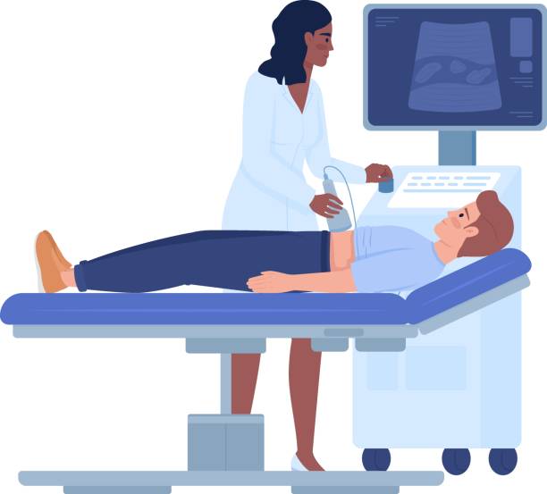
Most abdominal aortic aneurysms are asymptomatic and are found incidentally during routine imaging tests, such as an abdominal ultrasound, CT scan, or MRI. If your doctor suspects an AAA, they may order one or more of these tests to confirm the diagnosis.

In addition to imaging tests, your doctor may also perform a physical exam to check for the presence of an AAA. They will listen to your abdomen with a stethoscope to detect any abnormal sounds, and may also feel for a pulsating mass in the abdomen.

Visit our service page: Abdominal Aorta Aneurysm Scan

Fatty liver

Treatment of AAA

The treatment for an abdominal aortic aneurysm depends on several factors, including the size of the aneurysm, your age, and your overall health. Treatment options include:

Watchful waiting: If the aneurysm is small (less than 5 centimeters) and you have no symptoms, your doctor may recommend regular monitoring with imaging tests to watch for any growth or changes.

Endovascular repair: Endovascular repair is a minimally invasive procedure that involves placing a stent graft through a small incision in the groin to reinforce the weakened area of the aorta. This procedure is typically used for aneurysms that are large or growing quickly.

Open surgery: Open surgery involves removing the damaged portion of the aorta and replacing it with a synthetic graft. This procedure is typically used for larger aneurysms or when endovascular repair is not possible.

Medications: Your doctor may prescribe medications to control blood pressure, reduce cholesterol levels, or prevent blood clots from forming, which can help slow the growth of an AAA.

Conclusion

Abdominal aortic aneurysm is a serious condition that requires prompt diagnosis and treatment to prevent potentially life-threatening complications. If you are at risk for an AAA or have symptoms such as abdominal pain or swelling, it is important to talk to your doctor about getting screened with imaging tests such as an abdominal ultrasound. Early detection and treatment can improve outcomes and reduce the risk of rupture.

FAQs

What are the risk factors for abdominal aortic aneurysm?
The risk factors for AAA include age (over 60), male gender, smoking, high blood pressure, high cholesterol, and a family history of the condition.

Is there anything I can do to prevent an abdominal aortic aneurysm?
The best way to prevent an AAA is to avoid smoking, maintain a healthy weight, and manage conditions such as high blood pressure and high cholesterol.

What are the symptoms of an abdominal aortic aneurysm?
Most abdominal aortic aneurysms are asymptomatic, but some people may experience abdominal pain, back pain, or a pulsating mass in the abdomen.

How often should I get screened for abdominal aortic aneurysm?
The frequency of screening for AAA depends on your age, gender, and risk factors. Your doctor can recommend a screening schedule based on your individual risk profile.

Is surgery for an abdominal aortic aneurysm risky?
All surgeries carry some degree of risk, but the risk of complications from surgery for AAA is generally low. Your doctor will discuss the risks and benefits of each treatment option with you to help you make an informed decision.

What Is the Difference Between Pelvic Scan and Abdominal Scan?

Medical imaging is critical in detecting and assessing a variety of disorders affecting the abdominal and pelvic areas. The abdominal and pelvic scans are two regularly used scans in this area. While both scans concentrate on different parts of the body, they serve separate functions and give significant information to healthcare practitioners. In this blog article, we’ll look at the distinctions between a pelvic scan and an abdominal scan in order to better comprehend their functions in medical imaging.

Scan of the Abdomen

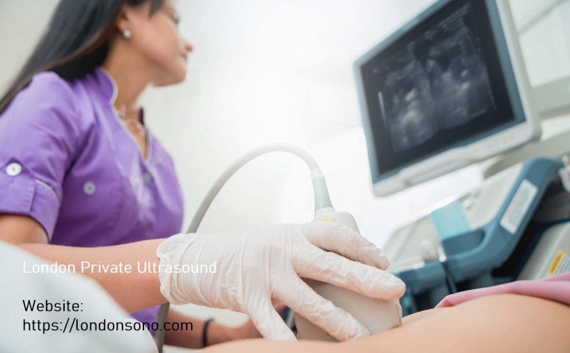
An abdominal scan, often known as an abdominal ultrasound, is a non-invasive imaging treatment that takes comprehensive pictures of the organs and structures in the belly. It uses high-frequency sound waves to generate real-time visuals on a display. A portable instrument called a transducer is softly pushed across the skin of the abdomen during an abdominal scan, producing sound waves and recording the returning echoes to make the pictures.

An abdominal scan’s primary goal is to assess the organs located in the abdominal cavity, which include the liver, gallbladder, pancreas, spleen, kidneys, and bladder, among others. It reveals important details on the size, shape, texture, and anomalies of these organs. A scan of the abdomen can aid in the diagnosis of illnesses such as liver disease, gallstones, pancreatic abnormalities, kidney stones, and abdominal tumours. It may also measure blood flow in the main arteries of the belly.

Scan of the Pelvic Organs

A pelvic scan, also known as a pelvic ultrasound, on the other hand, checks the organs and tissues in the pelvic area. The uterus, ovaries, fallopian tubes (in females), prostate gland (in men), bladder, and surrounding tissues are all part of the pelvic region. A pelvic scan, like an abdominal scan, uses sound waves and a transducer to produce real-time pictures.

A pelvic scan is used to assess the reproductive organs in both males and females. It aids in the diagnosis of disorders such as fibroids, ovarian cysts, polycystic ovary syndrome (PCOS), endometriosis, and pelvic inflammatory disease in females. It is also used during pregnancy to monitor foetal growth and assess the placenta. In men, a pelvic scan can detect problems such as benign prostatic hyperplasia (enlargement) or tumours in the prostate gland.

Distinctions and overlaps

The abdominal scan focuses on the organs of the abdominal cavity, whereas the pelvic scan focuses on the reproductive organs and associated tissues in the pelvic area. There is, however, some overlap between the two images. Both scans, for example, might look for abnormalities in the bladder, such as stones or tumours. To obtain a full examination of the abdominal and pelvic areas, a healthcare expert may propose a combination of both scans, known as a transabdominal and transvaginal ultrasound.

Finally, an abdominal scan and a pelvic scan are two independent imaging techniques that give important information about various parts of the body. The abdominal scan examines the organs in the abdominal cavity, whereas the pelvic scan examines the reproductive organs and associated tissues in the pelvic area. Both scans are non-invasive, safe, and often used to identify a wide range of disorders. If you have particular symptoms or concerns about your abdomen or pelvis, it is critical to contact with a healthcare specialist who can prescribe the best scan for your unique requirements.

Doctor is performing liver biopsy to a young women...supporting with Ultrasound
August 10, 2023 by Londonsono 0 Comments

Exploring Abdominal Ultrasound Scans:  A Non-Invasive Approach to Diagnosis

An abdominal ultrasound scan, a commonly employed diagnostic tool, offers a comprehensive view of key abdominal organs. This non-invasive procedure provides valuable information about the abdominal aorta, liver, gallbladder, pancreas, kidneys, and spleen. These information play a pivotal role in diagnosing or excluding various abdominal conditions that often lead to pain and discomfort in the upper abdomen, such as gallstones and kidney stones.

One of the most significant advantages of ultrasound is its painless and safe nature. This procedure entails no adverse effects, either during or after the examination, this making it a preferred choice for many individuals seeking medical insights.

Within the realm of liver health, ultrasound plays a vital role in identifying hepatic steatosis, commonly referred to as fatty liver. This condition involves the accumulation of fatty deposits in the liver. While a certain amount of fat in the liver is normal, a fat content exceeding 5 to 10 percent classifies as fatty liver disease. Importantly, this condition is reversible and can be managed through diet and exercise.

Non-alcoholic fatty liver disease (NAFLD) is another common disorder, characterized by fat accumulation in the liver among individuals who consume minimal or no alcohol. The most frequent manifestation of NAFLD is fatty liver, a non-serious condition.

Addressing fatty liver entails a multifaceted approach, including safe weight loss, triglyceride reduction through dietary changes or medication, abstaining from alcohol, diabetes management if applicable, a balanced diet, and increased physical activity.

Ultrasound scans are indeed capable of detecting cancer within the abdominal organs. An abdominal ultrasound assesses the liver, kidneys, pancreas, gallbladder, and spleen for any abnormalities. If a mass indicative of possible abdominal cancer is observed during ultrasound, further imaging through CT or MRI is required to confirm its existence.

The duration for scheduling an abdominal ultrasound varies across NHS hospitals, typically taking place within approximately six weeks. In private clinics, like ours, it’s possible to secure a same-day or next-day appointment.

Contrary to inaccurate online information, fasting for an abdominal ultrasound doesn’t necessitate an excessive 8 to 12 hours. Merely fasting for four hours suffices, and you can consume your medication with clear fluids like water during this period.

The benefits of abdominal ultrasound are manifold: it’s non-invasive, painless, widely available, cost-effective, and doesn’t employ ionizing radiation. However, ultrasound has limitations; it struggles to penetrate bowel or gas, hindering the evaluation of abdominal organs obscured by these factors. Also, visualizing internal anatomy in larger individuals can be challenging due to the greater distance sound waves need to travel.

Abdominal scan results are interpreted by a highly trained ultrasonographer. They perform the ultrasound, provide a diagnostic report, and discuss the findings with you during and after the examination.

At our clinic, located in the heart of London, we offer same-day appointments in a compassionate environment. Our experienced, qualified sonographers are registered with the HCPC, BMUS, and SoR, ensuring your health is in capable hands.

Choosing us for your private abdominal ultrasound scan means benefiting from our convenient location, same-day appointments, and the expertise of our qualified sonographers. We prioritize affordability without compromising diagnostic accuracy or service quality, offering competitive prices and transparency.

If you seek an abdominal ultrasound scan in London, you’re at the right place. At London Private Ultrasound, we offer a range of ultrasound scans, conveniently located in central London. Our consultant sonographers, experienced in medical ultrasound, guarantee exceptional service quality and diagnostic accuracy. We offer flexible same-day and out-of-hours appointments, ensuring your convenience.

Inquiring about the cost of private abdominal scans? Our philosophy revolves around affordable healthcare, ensuring ultrasound imaging remains accessible to all. Our pricing is competitive and transparent, with no hidden costs.

Booking an abdominal ultrasound is simple through our online booking diary, offering evening and weekend appointments. If you face any issues, feel free to reach out via email or phone.

Abdominal ultrasound, also known as liver ultrasound, is a diagnostic investigation that assesses your liver for abnormalities. Unlike CT scans, ultrasound uses sound waves instead of radiation.

The benefits of abdominal ultrasound are abundant, including non-invasiveness, painlessness, accessibility, cost-effectiveness, and the absence of ionizing radiation. No harmful effects on humans are associated with ultrasound examinations.

While ultrasound is powerful, it does have limitations. It struggles to penetrate bowel or gas, limiting the evaluation of abdominal organs. Additionally, visualizing internal anatomy in larger individuals can be challenging.

In conclusion, abdominal ultrasound offers valuable insights into abdominal health, covering a range of vital organs. This non-invasive, painless, and accessible procedure aids in diagnosing and managing various conditions, and is particularly significant in assessing liver health.