27 Welbeck Street, London W1G 8EN
Tel: 020 7101 3377

Category: Vascular Ultrasound Scans

Vascular ultrasound scans, a specialized form of medical ultrasound, focus on assessing blood flow within arteries and veins. These non-invasive exams employ high-frequency sound waves to create detailed images of the vascular system. They are vital for diagnosing conditions like deep vein thrombosis, arterial blockages, and aneurysms, helping healthcare professionals plan appropriate treatments and interventions for improved cardiovascular health.
September 3, 2023 by London Private Ultrasound 0 Comments

Why DVT Ultrasound Scans are Essential for Accurate Diagnosis

Introduction:

Deep Vein Thrombosis (DVT) is a serious medical condition that occurs when a blood clot forms in a deep vein, often in the legs. It can lead to severe complications if left untreated, including pulmonary embolism, which can be fatal. In order to accurately diagnose DVT, healthcare professionals rely on various imaging techniques, with ultrasound playing a crucial role. This article will explore why DVT ultrasound scans are essential for accurate diagnosis, highlighting their benefits, the procedure, and their role in preventing potential complications.

Understanding DVT and Its Risks:

Before delving into the importance of ultrasound scans, it is important to grasp the nature of DVT and its associated risks. DVT primarily affects the deep veins in the legs, where blood flow is slower compared to the arteries. This sluggish blood flow, combined with certain risk factors such as prolonged immobility, obesity, and family history, increases the likelihood of blood clot formation. If a clot dislodges and travels to the lungs, it can cause a life-threatening pulmonary embolism.

The Role of Ultrasound in DVT Diagnosis:

Ultrasound is a non-invasive imaging technique that utilizes sound waves to produce real-time images of the body’s internal structures. In the case of DVT, ultrasound plays a vital role in diagnosing the condition accurately. It allows healthcare professionals to visualize the veins, identify blood clots, and assess their extent and location. Unlike other imaging methods like magnetic resonance imaging (MRI) or computed tomography (CT) scans, ultrasound does not involve ionizing radiation, making it safer for patients.

Advantages of DVT Ultrasound Scans:

  1. Accuracy: DVT ultrasound scans are known for their high accuracy in detecting blood clots. They provide real-time images, enabling immediate visual confirmation of DVT, thereby facilitating prompt treatment initiation.

  2. Safety: As mentioned earlier, ultrasound scans do not expose the patient to any harmful radiation. This makes it a safer option, particularly for pregnant women, children, and individuals who require repeated scans.

  3. Non-invasiveness: Unlike other diagnostic procedures, such as venography, which involves injecting a contrast dye into the veins, ultrasound scans are non-invasive. It involves the use of a handheld device or transducer that is gently moved over the skin.

  4. Cost-effectiveness: DVT ultrasound scans are generally more affordable compared to other imaging techniques, making them accessible to a wider range of patients. This affordability allows for more frequent monitoring of patients at risk and facilitates early detection.

The DVT Ultrasound Procedure:

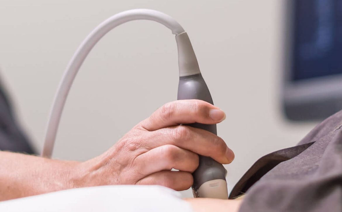
During a DVT ultrasound scan, a healthcare professional applies a gel to the skin over the area being examined. The transducer, which emits sound waves, is then placed on the gel-coated skin. The healthcare professional moves the transducer along the veins, capturing images that are displayed on a monitor in real-time. By analyzing the images, they can identify any blood clots present and assess their severity. The procedure is painless and typically takes less than 30 minutes to complete.

Preventing Complications and Ensuring Proper Treatment:

Early and accurate diagnosis of DVT through ultrasound scans is crucial for initiating appropriate treatment promptly. Anticoagulant medications, also known as blood thinners, are commonly prescribed to prevent further clot formation and reduce the risk of pulmonary embolism. Depending on the severity of the clot, more aggressive treatments like thrombolytic therapy may be considered.

Conclusion:

DVT ultrasound scans play a fundamental role in accurately diagnosing DVT, allowing for timely treatment and reducing the risk of life-threatening complications. The advantages of ultrasound, including its safety, non-invasiveness, cost-effectiveness, and high accuracy, make it the preferred diagnostic option for healthcare professionals. By utilizing ultrasound technology, physicians can closely monitor patients at risk, detect DVT at its earliest stages, and provide appropriate care.

September 3, 2023 by London Private Ultrasound 0 Comments

5 Vital Reasons Why You Should Consider a Carotid Doppler Ultrasound

Carotid Doppler - cardiovascular health

When it comes to your cardiovascular health, knowledge is power. Carotid Doppler screening via ultrasound is a non-invasive diagnostic test that can provide valuable insights into your vascular well-being. In this article, we will explore the five vital reasons why you should consider a Carotid Doppler scan. Discover how the carotid doppler can help you maintain optimal health and prevent potentially life-threatening conditions.

Why a carotid doppler?

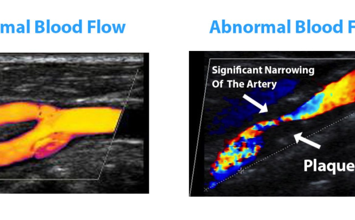
  1. Early Detection of Carotid Artery Disease: Carotid artery disease occurs when fatty deposits, known as plaque, build up in the arteries that supply blood to the brain. This condition can increase the risk of stroke. By undergoing a Carotid Doppler Ultrasound, you can detect any blockages or abnormalities at an early stage. Early detection allows for prompt preventive measures and reduces the risk of stroke or other cardiovascular complications.

  2. Assessing Stroke Risk: Comprehending your individual stroke risk is essential in managing your overall health. A Carotid Doppler Ultrasound helps evaluate the narrowing of your carotid arteries, indicating if you are at an increased risk of stroke. Armed with this knowledge, you can work together with your healthcare provider to implement lifestyle changes or medical interventions to minimize this risk effectively. Learn more with our cholesterol health check-up and diabetes health-chck up designed to help you manage your cholesterol and lower your risk of stroke.

  3. Monitoring Blood Flow to the Brain: The brain relies on a steady supply of oxygen-rich blood to function optimally. A Carotid Doppler Ultrasound enables healthcare professionals to assess the blood flow in real-time and identify any abnormalities or blockages. Understanding the efficiency of blood flow to the brain ensures early intervention, minimizing the chances of cognitive decline or other related conditions.

  4. Identification of Silent Strokes: Silent strokes, also known as silent cerebral infarcts, have no noticeable symptoms. These mini-strokes can cause brain damage and contribute to cognitive decline over time. By undergoing a Carotid Doppler Ultrasound, healthcare professionals can detect any silent strokes that may have occurred, allowing for early intervention and minimizing the potential long-term impact on brain health.

  5. Personalized Treatment and Prevention Planning: The Carotid Doppler Ultrasound provides valuable information about your vascular health, allowing healthcare providers to tailor a personalized treatment and prevention plan. This diagnostic test helps identify individuals who may benefit from lifestyle modifications, medication, or medical procedures to maintain a healthy cardiovascular system. By following the recommendations derived from this test, you can actively play a role in reducing your risk of stroke and other cardiovascular diseases.

London's Private Vascular services

Choosing Carotid Doppler Screening can have a significant impact on your overall health and well-being. With the ability to detect early signs of carotid artery disease, evaluate stroke risk, monitor blood flow, identify silent strokes, and personalise treatment plans, this non-invasive test is an essential tool in maintaining a healthy cardiovascular system. Take charge of your health today and consider a Carotid Doppler Ultrasound. 

No referral is needed and you can book an appointment with a private doctor at our central London clinic or opt for a pre-appointment online consultation.

London Private Ultrasound provide a full suite of Vascular services and have world renowned medical experts able to advise and guide you through the options, providing you with highly specific packages such as the Carotid Doppler Screening or specialist scans for blood clots causing Deep Vein Thrombosis or an Abdominal Aorta Aeurysm scan

The cardiovascular system is fundamental to achieving optimal health and as well as providing scans for those experiencing symptoms and difficulties we also provide an extensive range of preventative health checks – these designed to identify potential warning signs and allow us to prescribe preventative measures to stem the possibilities of conditions or diseases developing.

12 Health Screenings Women Should Have

Essential Screenings for Women's Health

Taking care of your health as a woman is critical to live a happy and healthy life. Regular health checkups are one approach to ensure your general well-being. You can take proactive efforts to avoid or control possible health concerns if you notice them early on. This essay will go over 12 important health tests that every woman should get.

Pap Smear A Pap smear is a screening test for cervical cancer. Women should start obtaining Pap smears at the age of 21 and get one every three years until they are 65.

Mammogram
A mammogram is a low-dose X-ray that is used to detect breast cancer. Mammograms should begin at the age of 40 and be repeated every one to two years.

Bone Density Exam
A bone density test is used to diagnose osteoporosis, a disorder in which bones become weak and brittle. Every two years, women over the age of 65 should get a bone density test.

Blood Pressure Check
High blood pressure can cause cardiovascular disease and stroke. Women’s blood pressure should be monitored at least once a year.

Test for Cholesterol
High cholesterol levels can contribute to cardiovascular disease. Beginning at the age of 20, women should have their cholesterol levels examined every four to six years.

Screening for Skin Cancer
In the United States, skin cancer is the most frequent kind of cancer. Every year, women should get their skin evaluated by a dermatologist.

Please see our service page: Women’s Health Examination

women health

Colon Cancer Screening

Colon cancer is the second leading cause of cancer deaths in women. Women should begin getting screened at age 50 or earlier if they have a family history of colon cancer.

Thyroid Test

The thyroid gland regulates metabolism. Women should have their thyroid checked every five years, starting at age 35.

Diabetes Screening

Diabetes can lead to heart disease, stroke, and other complications. Women should get screened for diabetes every three years starting at age 45.

STD Testing

Sexually transmitted diseases (STDs) can cause serious health problems. Women who are sexually active should get tested for STDs at least once a year.

Eye Exam

Regular eye exams can detect eye diseases such as glaucoma and cataracts. Women should have an eye exam every two years.

Mental Health Screening

Mental health is just as important as physical health. Women should get screened for depression and anxiety regularly.

Womens Health Screening Packages

Womens Health Packages

In conclusion, women’s health screenings are an essential part of maintaining good health. By getting regular health check-ups, you can detect potential health problems early on and take proactive steps to prevent or manage them. If you have any concerns about your health, be sure to talk to your healthcare provider about which screenings are right for you.

London Private Ultrasound are an established Ultrasound Clinic in Central London providing specialist Ultrasound services and expert clinicians in a range of health issues, this to allow you to have the expertise required to achieve optimal health.


Shedding Light on Peripheral Artery Disease: Understanding Doppler Ultrasound

Peripheral Artery Disease (PAD) is a condition that affects blood flow to the limbs, most commonly the legs. It can lead to pain, discomfort, and even serious complications if left untreated. Doppler ultrasound is a crucial diagnostic tool used to assess blood circulation in the extremities and diagnose PAD. In this blog post, we will explore the significance of Doppler ultrasound in diagnosing PAD, how the procedure works, its benefits, and its role in improving patient outcomes.

The Role of Doppler Ultrasound in PAD Diagnosis

Doppler ultrasound is a non-invasive imaging technique that uses sound waves to evaluate blood flow and pressure within blood vessels. When applied to PAD diagnosis, Doppler ultrasound assists in:

  1. Blood Flow Assessment: Doppler ultrasound detects and measures the speed and direction of blood flow in arteries and veins. This information helps healthcare providers identify blockages, narrowing, or restrictions in blood vessels—common characteristics of PAD.

  2. Ankle-Brachial Index (ABI) Calculation: ABI is a key indicator of PAD severity. It’s determined by comparing blood pressure measurements at the ankle and the arm. Doppler ultrasound is essential in obtaining accurate blood pressure measurements at these locations to calculate the ABI.

  3. Identifying Stenosis and Occlusions: Doppler ultrasound’s ability to visualize blood flow allows healthcare professionals to identify stenosis (narrowing) and occlusions (blockages) within blood vessels. This information is crucial for treatment planning.

  4. Monitoring Disease Progression: Doppler ultrasound is used to monitor the progression of PAD over time. Regular ultrasounds can help healthcare providers assess the effectiveness of treatment and make necessary adjustments.

The Doppler Ultrasound Procedure for PAD Diagnosis

  1. Preparation: The procedure generally doesn’t require extensive preparation. You might be asked to wear loose clothing or change into a gown for easier access to the limbs.

  2. Procedure: During the ultrasound, a gel is applied to the skin over the area being examined. The gel helps the sound waves travel smoothly and prevents air gaps. The ultrasound technician uses a handheld device called a transducer, which emits sound waves and captures the echoes produced by the movement of blood. These echoes are translated into visual and auditory information.

  3. Blood Flow Assessment: The technician will focus the transducer on different areas of the limb, including the arteries and veins. By analyzing the sound waves, the technician can create images and measurements of blood flow.

  4. ABI Calculation: The technician will use a blood pressure cuff and Doppler ultrasound to measure blood pressure at the arm and ankle. These measurements are used to calculate the ABI, a key parameter in diagnosing PAD.

Benefits of Doppler Ultrasound for PAD

  1. Non-Invasive: Doppler ultrasound is non-invasive, meaning it doesn’t require surgical procedures or injections.

  2. Accurate Diagnosis: Doppler ultrasound provides real-time visualizations of blood flow, aiding in accurate diagnosis and assessment.

  3. Early Detection: Early detection of PAD through Doppler ultrasound allows for timely intervention and better management of the condition.

  4. Risk Assessment: ABI calculation using Doppler ultrasound helps assess the risk of complications such as ulcers and amputations.

Conclusion

Doppler ultrasound plays a pivotal role in diagnosing and managing Peripheral Artery Disease. By providing detailed insights into blood flow and pressure, it enables healthcare providers to make informed decisions about treatment and lifestyle changes. If you’re experiencing symptoms such as leg pain, cramping, or numbness, consulting a medical professional and considering a Doppler ultrasound can contribute to preserving your vascular health and overall well-being.