27 Welbeck Street, London W1G 8EN
Tel: 020 7101 3377

Category: Musculoskeletal Ultrasound Scan

Musculoskeletal ultrasound scans are diagnostic procedures that employ high-frequency sound waves to produce real-time images of the musculoskeletal system. These scans are particularly valuable for assessing soft tissues, tendons, ligaments, muscles, and joints. They help in diagnosing a wide range of musculoskeletal conditions such as tendonitis, ligament injuries, arthritis, and joint abnormalities.
September 5, 2023 by London Private Ultrasound 0 Comments

Diagnosing Elbow related issues with Ultrasound Scans

Elbow injuries can be particularly debilitating, affecting people in various age groups and professions. Thankfully, with advancements in medical technology, elbow ultrasound scans have emerged as a non-invasive and effective diagnostic tool. In this article, we will explore the advantages of elbow ultrasound scans and shed light on how they can help in diagnosing and managing elbow-related conditions.

Understanding Elbow Ultrasound Scans:

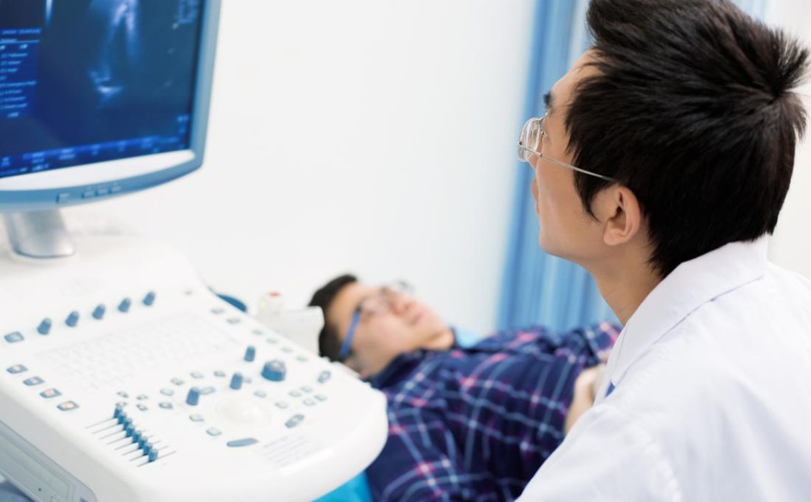
Elbow ultrasound scans utilize high-frequency sound waves to create detailed images of the internal structures of the elbow joint. This painless procedure is performed by a skilled sonographer or a qualified radiologist. By producing real-time images, elbow ultrasound scans provide valuable insights into potential abnormalities, assisting in accurate diagnosis and efficient treatment planning.

Advantages of Elbow Ultrasound Scans:

  1. Non-Invasive and Safe: Elbow ultrasound scans are non-invasive, meaning they do not require any surgical incisions. Unlike other imaging techniques such as X-rays or CT scans, ultrasound scans do not use ionizing radiation, making them safe for repeat examinations, even for pregnant women and children.

  2. Real-Time Imaging: One significant advantage of elbow ultrasound scans is the ability to observe the elbow joint in real-time. This feature allows healthcare professionals to evaluate joint mobility, identify abnormalities during movement, and assess the response to specific maneuvers or treatments.

  3. Detailed Visualization: Elbow ultrasound scans provide detailed images of the muscles, tendons, ligaments, and other soft tissues of the elbow joint. This level of visualization helps in identifying various conditions, including tendon tears, bursitis, arthritis, and inflammation, leading to more accurate diagnoses.

  4. Guided Procedures: Elbow ultrasound scans can also be used as a guide during various minimally invasive procedures, such as injections or aspirations. The real-time imaging capability assists physicians in precisely targeting the affected area, ensuring the delivery of medications or fluid removal with increased accuracy.

  5. Cost-Effective: Compared to other imaging modalities like MRI or CT scans, elbow ultrasound scans are generally more cost-effective. They offer a viable option for initial evaluation, enabling healthcare providers to assess the condition before recommending further investigative procedures, potentially saving both time and money for the patient.

Conclusion:

In conclusion, elbow ultrasound scans offer numerous advantages in diagnosing and managing elbow-related conditions. With their non-invasive nature, real-time imaging capabilities, and ability to visualize soft tissue structures, these scans have become an indispensable tool for healthcare professionals. By considering the advantages discussed in this article, individuals experiencing elbow pain or injuries can seek appropriate medical attention, leading to accurate diagnoses and effective treatment plans.

September 3, 2023 by London Private Ultrasound 0 Comments

Unveiling the Hidden Truth: Lumps and Bumps Ultrasound Scan

Introduction:

When it comes to medical diagnostic imaging, ultrasound scans have become an integral tool in the detection and evaluation of various conditions. Among these, the identification and examination of lumps and bumps through ultrasound have proven to be highly effective. In this article, we will delve into the hidden truths of lumps and bumps ultrasound scans, demystifying the process and shedding light on its significance in diagnosing underlying health concerns.

Understanding Lumps and Bumps Ultrasound Scans:

Ultrasound scans, also known as sonograms, utilize high-frequency sound waves to create images of the internal structures of the body. These scans offer a non-invasive and safe approach to visualize lumps, bumps, or masses, providing crucial information about their nature and characteristics.

  1. The Importance of Lumps and Bumps Ultrasound Scans:

Lumps and bumps can evoke fear and uncertainty in individuals, often causing anxiety about potential health issues. Ultrasound scans are particularly valuable in reassuring patients or identifying potential concerns, as they can differentiate between solid masses, fluid-filled cysts, or benign tumors. With early detection being vital in many cases, ultrasound scans offer a proactive approach to diagnosis and subsequent treatment.

  1. Indications and Applications:

Lumps and bumps ultrasound scans are used across various medical specialties, serving as a primary diagnostic tool or guiding further explorations. Some common indications for these scans include:

a. Breast Abnormalities: Ultrasound scans help determine the nature of breast lumps or abnormalities detected during self-examinations or mammograms. This method aids in differentiating between benign cysts and potentially malignant masses.

b. Thyroid Nodules: The evaluation of thyroid nodules is another significant use of ultrasound scans. By assessing the size, shape, and internal characteristics of nodules, doctors can decide whether further interventions, such as biopsies, are necessary.

c. Musculoskeletal Lumps: For individuals with palpable lumps or bumps in their muscles or joints, ultrasound scans can provide valuable information about the structures involved, aiding in accurate diagnoses and treatment planning.

d. Abdominal Masses: Ultrasound scans can detect and characterize lumps or masses in the abdominal region, assisting in differentiating between solid tumors, cysts, or other anomalies.

  1. The Ultrasound Process and Techniques:

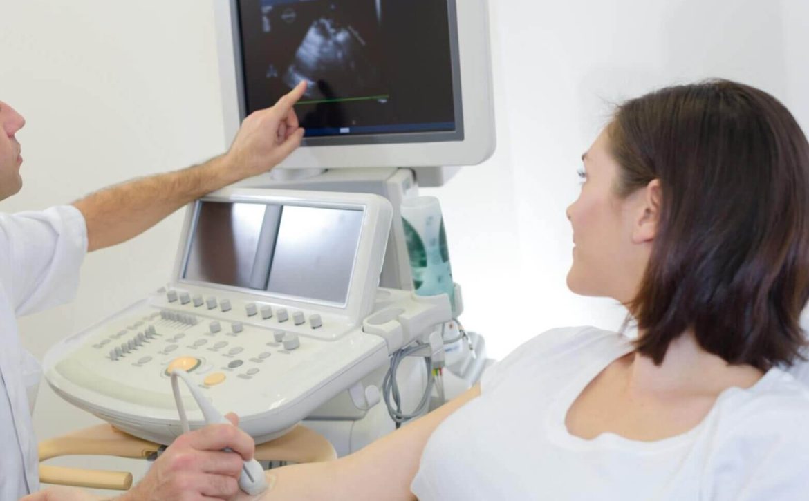
During a lumps and bumps ultrasound scan, a skilled sonographer or radiologist utilizes a handheld transducer to send and receive sound waves. The transducer is applied to the skin, coupled with a gel that enhances sound wave transmission. The produced images are then analyzed, interpreted, and communicated to the patient by a medical professional.

a. Real-Time Imaging: One of the significant advantages of ultrasound scans is that they offer real-time imaging. This means that the live images allow for dynamic assessments, ensuring a comprehensive evaluation of lumps and bumps.

b. Doppler Ultrasound: In certain cases, when assessing blood flow patterns within lumps or evaluating vascular abnormalities, a Doppler ultrasound technique may be employed. This technique provides additional insights into the vascularity and blood supply of the detected lesions.

Conclusion:

In conclusion, lumps and bumps ultrasound scans provide an invaluable tool in detecting, characterizing, and evaluating various conditions across different medical specialties. These scans help alleviate patient concerns by providing accurate information about the nature and composition of lumps or bumps. By encouraging early intervention and facilitating timely treatment, ultrasound scans play a crucial role in maintaining and promoting overall health.

September 2, 2023 by London Private Ultrasound 0 Comments

The Advantages of Elbow Ultrasound Scans Revealed

Introduction: Elbow injuries and conditions can significantly impact an individual’s mobility and overall quality of life. In such cases, accurate diagnosis becomes crucial for effective treatment. While there are various diagnostic imaging techniques available, including X-rays and MRI scans, elbow ultrasound scans have emerged as a reliable and advantageous option. In this article, we will delve into the numerous benefits of elbow ultrasound scans and shed light on why they are considered a valuable diagnostic tool in the medical field.

What is Elbow Ultrasound? Elbow ultrasound, also known as sonography or ultrasonography, employs high-frequency sound waves to create detailed images of the internal structures of the elbow joint. By using a handheld transducer and a gel that helps transmit sound waves, medical professionals can visualize the bones, muscles, tendons, and ligaments in real-time.

Benefits of Elbow Ultrasound Scans:

  1. Non-invasive and Safe: Unlike other imaging techniques, such as X-rays or CT scans, elbow ultrasound scans do not expose patients to harmful ionizing radiation. This makes them a safer option, especially for individuals who may require repeated imaging or those more susceptible to radiation-related risks, such as children and pregnant women.

  2. Real-time Visualization: One of the significant advantages of elbow ultrasound scans is the ability to visualize the elbow joint in real-time. Medical professionals can observe the structures while they are in motion, providing valuable insights into any abnormalities or dysfunctional movements. This dynamic visualization aids in diagnosing various conditions accurately and monitoring treatment progress.

  3. High Precision and Detail: With advanced technology, elbow ultrasound scans have become increasingly accurate and detailed. They provide exceptional resolution, allowing clinicians to identify even subtle abnormalities, such as tears in tendons, inflammation, or fluid accumulation. The detailed images obtained contribute to precise diagnosis and guide appropriate treatment plans.

  4. Cost-effective: Compared to other imaging modalities like MRI or CT scans, elbow ultrasound scans are relatively more affordable. Additionally, they can be performed in clinics or medical facilities, eliminating the need for hospital visits or extended stays. The cost-effectiveness of elbow ultrasounds makes them accessible to a wider range of patients, streamlining the diagnostic process.

  5. No Special Preparations Needed: Unlike some imaging techniques, elbow ultrasound scans require minimal preparations. Patients do not need to fast or undergo any special dietary restrictions before the procedure. This convenience adds to the overall patient experience and eliminates any unnecessary anxiety or inconvenience associated with complex preparations.

  6. Versatile and Comprehensive: Elbow ultrasound scans can provide a comprehensive assessment of various conditions, including but not limited to:

  • Tennis or Golfer’s elbow
  • Elbow bursitis
  • Ligament and tendon tears
  • Arthritis
  • Tumors or cysts within the elbow joint
  • Inflammation or fluid accumulation

By obtaining a clear and holistic view of the affected area, medical professionals can devise appropriate treatment plans tailored to the specific condition.

Conclusion: Elbow ultrasound scans have revolutionized the field of diagnostic imaging by offering several advantages over traditional methods. The non-invasive nature, real-time visualization, high precision, cost-effectiveness, and versatility make them a valuable tool in diagnosing and monitoring elbow conditions. With their ability to provide detailed and accurate imaging, elbow ultrasound scans have become a crucial component in the armamentarium of medical professionals, optimizing patient care and facilitating effective treatment plans. If you are experiencing elbow pain or suspect an injury, consult with a healthcare provider who can determine if an ultrasound scan is appropriate for your case.

September 1, 2023 by London Private Ultrasound 0 Comments

Everything You Need to Know about Lumps and Bumps Ultrasound Scan

Introduction:

Lumps and bumps can often be a cause for concern, causing anxiety and worry. In order to properly diagnose and address these issues, medical professionals often recommend an ultrasound scan. This non-invasive procedure has proven to be an effective way to identify and assess lumps and bumps, aiding in early detection and accurate diagnosis. In this article, we will delve into the world of lumps and bumps ultrasound scan, exploring its benefits, procedure, and what you can expect during your appointment.

Understanding Lumps and Bumps Ultrasound Scan:

A lumps and bumps ultrasound scan is a medical imaging technique that uses sound waves to create detailed pictures of the inside of your body. Unlike X-rays or CT scans, ultrasound scans do not involve the use of radiation, making them a safer option, especially for pregnant women or children. This procedure is commonly used to examine various areas of the body, including the breasts, neck, abdomen, and limbs.

Procedure:

Before the scan, your sonographer will explain the process to you and answer any questions you may have. You will be asked to lie down on an examination table, and a gel will be applied to the area being scanned. The sonographer will then use a small handheld device called a transducer to pass over the gel-covered area. This device emits high-frequency sound waves, which bounce back and create images on a monitor. The entire process is painless and typically takes around 15 to 30 minutes, depending on the area being examined.

Benefits of Lumps and Bumps Ultrasound Scan:

  1. Non-invasive: Unlike other imaging techniques, ultrasound scans are non-invasive, meaning they do not require any incisions or injections. This makes them a preferred choice for patients who prefer a less invasive approach.

  2. Safe and radiation-free: As mentioned earlier, ultrasound scans do not involve the use of radiation, making them safe for individuals of all ages, including pregnant women and children.

  3. Accurate and detailed imaging: Ultrasound scans provide real-time images of the area being examined, allowing the sonographer to view the internal structures and identify any abnormalities accurately. This helps in the early detection of potential health issues.

  4. Versatility: Lumps and bumps ultrasound scans can be used to examine various parts of the body, making them a versatile tool in the field of diagnostic medicine.

Determining Lumps and Bumps Differences with a Scan

  1. Breast examination: Ultrasound scans play a crucial role in the evaluation of breast lumps. It helps in distinguishing between lump and bump differences of solid masses and fluid-filled cysts, aiding in the diagnosis of breast cancer.

  2. Thyroid evaluation: Ultrasound scans are often used to assess the thyroid gland for any abnormalities, again this Thyroid ultrasound allows us to identify the lump and bump difference that occurs in nodules or cysts, for example.

  3. Abdominal evaluation: To investigate abdominal pain or detect abnormalities in the liver, gallbladder, pancreas, or kidneys, ultrasound scans are commonly recommended.

  4. Musculoskeletal examination: Ultrasound scans can be used to evaluate various musculoskeletal conditions, such as tendonitis, muscle tears, and joint inflammation. The causes will show up as a lump and bump difference in the mass type, density and manoeuvrability.

Monitor different types of Lumps and Bumps with ultrasound

Obtaining a lumps and bumps ultrasound scan with London Private Ultrasound is a valuable tool in the diagnosis and monitoring of various health conditions. Its non-invasive nature, safety, and versatility make it an ideal choice for patients seeking a detailed and accurate evaluation of lumps and bumps.

If you notice any abnormalities or have concerns about lumps or bumps on your body, it is essential to take action and you are welcome to opt for one of the many ultrasound health scans and lump and bump scans we provide. Alternatively, you may prefer to book a consultation with us first – this can be via an online channel or in our specialist ultrasound clinic where we conduct ultrasounds throughout each and every day with specialised clinics for each type of medical issue. If you are wanting to find out more about what syptoms and signs may indicate cancer we can offer you the support and consultations alongside these ultrasound services.

September 1, 2023 by London Private Ultrasound 0 Comments

Why an Elbow Ultrasound Scan is Vital for Diagnosis

In the world of medical diagnostics, advancements in technology have played a significant role in improving accuracy and efficiency. One such technology that has revolutionized the field is the elbow ultrasound scan. By harnessing the power of sound waves, this non-invasive procedure has become a vital tool for diagnosing various conditions affecting the elbow joint.

So, why is an elbow ultrasound scan so important in the diagnosis process? Let’s explore its benefits and the role it plays in identifying and treating elbow-related conditions.

First and foremost, an elbow ultrasound scan provides healthcare professionals with a detailed image of the elbow joint’s internal structures. By using high-frequency sound waves, the scan captures real-time images, allowing for a comprehensive examination of the joint’s anatomy. This imaging technique helps in detecting abnormalities, fluid accumulation, growths, or any other structural damages that may be affecting the elbow joint.

One of the primary advantages of an ultrasound scan is its ability to provide real-time imaging. Unlike other imaging techniques, such as X-rays or magnetic resonance imaging (MRI), which may require additional time for development or interpretation, ultrasound produces immediate results. The real-time images allow healthcare professionals to visualize the elbow joint in motion, evaluating its functionality and detecting any abnormalities that may only occur during specific movements or positions.

Furthermore, an elbow ultrasound scan is a non-invasive procedure that eliminates the need for surgical intervention. Traditionally, obtaining an accurate diagnosis for elbow conditions required invasive procedures or exploratory surgeries, which are not only costly but also pose certain risks and complications. With the advent of ultrasound technology, healthcare professionals can now perform a thorough assessment of the elbow joint without subjecting the patient to unnecessary invasive procedures.

Another significant advantage of an elbow ultrasound scan is its cost-effectiveness. Compared to other imaging techniques, such as MRI or computed tomography (CT) scans, ultrasound scans are relatively more affordable. This affordability makes it accessible to a broader range of patients, ensuring that everyone has the opportunity to receive a timely and accurate diagnosis for their elbow-related conditions.

Now, let’s take a closer look at some specific elbow conditions that greatly benefit from an ultrasound scan.

Tennis elbow, medically known as lateral epicondylitis, is a common overuse injury affecting the tendons in the elbow region. An ultrasound scan helps in visualizing the inflamed or damaged tendons, allowing for proper identification and targeted treatment. It also assists in differentiating tennis elbow from other similar conditions, such as golfer’s elbow or radial tunnel syndrome.

Golfer’s elbow, or medial epicondylitis, is another condition that can be effectively diagnosed using an elbow ultrasound scan. By capturing real-time images of the affected area, the scan helps in assessing the extent of the injury, identifying any tendon abnormalities, and guiding appropriate treatment options.

In addition to diagnosing tendon-related conditions, an elbow ultrasound scan is also vital in detecting joint-related issues. Conditions such as arthritis, bursitis, or synovitis can be accurately diagnosed by evaluating the joint space, identifying any erosions or fluid accumulation, and determining the overall health and functionality of the joint.

In conclusion, an elbow ultrasound scan has revolutionized the diagnosis and treatment of various elbow-related conditions. This non-invasive and cost-effective imaging technique provides healthcare professionals with detailed and real-time images of the elbow joint, allowing for accurate identification of structural damages, abnormalities, or fluid accumulation. It plays a crucial role in diagnosing conditions such as tennis elbow, golfer’s elbow, arthritis, bursitis, or synovitis. By incorporating an elbow ultrasound scan into the diagnostic process, healthcare professionals can offer more targeted and effective treatment options to their patients.

12 Health Screenings Women Should Have

Essential Screenings for Women's Health

Taking care of your health as a woman is critical to live a happy and healthy life. Regular health checkups are one approach to ensure your general well-being. You can take proactive efforts to avoid or control possible health concerns if you notice them early on. This essay will go over 12 important health tests that every woman should get.

Pap Smear A Pap smear is a screening test for cervical cancer. Women should start obtaining Pap smears at the age of 21 and get one every three years until they are 65.

Mammogram
A mammogram is a low-dose X-ray that is used to detect breast cancer. Mammograms should begin at the age of 40 and be repeated every one to two years.

Bone Density Exam
A bone density test is used to diagnose osteoporosis, a disorder in which bones become weak and brittle. Every two years, women over the age of 65 should get a bone density test.

Blood Pressure Check
High blood pressure can cause cardiovascular disease and stroke. Women’s blood pressure should be monitored at least once a year.

Test for Cholesterol
High cholesterol levels can contribute to cardiovascular disease. Beginning at the age of 20, women should have their cholesterol levels examined every four to six years.

Screening for Skin Cancer
In the United States, skin cancer is the most frequent kind of cancer. Every year, women should get their skin evaluated by a dermatologist.

Please see our service page: Women’s Health Examination

women health

Colon Cancer Screening

Colon cancer is the second leading cause of cancer deaths in women. Women should begin getting screened at age 50 or earlier if they have a family history of colon cancer.

Thyroid Test

The thyroid gland regulates metabolism. Women should have their thyroid checked every five years, starting at age 35.

Diabetes Screening

Diabetes can lead to heart disease, stroke, and other complications. Women should get screened for diabetes every three years starting at age 45.

STD Testing

Sexually transmitted diseases (STDs) can cause serious health problems. Women who are sexually active should get tested for STDs at least once a year.

Eye Exam

Regular eye exams can detect eye diseases such as glaucoma and cataracts. Women should have an eye exam every two years.

Mental Health Screening

Mental health is just as important as physical health. Women should get screened for depression and anxiety regularly.

Womens Health Screening Packages

Womens Health Packages

In conclusion, women’s health screenings are an essential part of maintaining good health. By getting regular health check-ups, you can detect potential health problems early on and take proactive steps to prevent or manage them. If you have any concerns about your health, be sure to talk to your healthcare provider about which screenings are right for you.

London Private Ultrasound are an established Ultrasound Clinic in Central London providing specialist Ultrasound services and expert clinicians in a range of health issues, this to allow you to have the expertise required to achieve optimal health.


Upper Limb Ultrasound Scan

Ultrasonography has impaled through various medical specialities as a multipurpose imaging tool for assessment and evaluation of superficial and deep structures. It has been introduced to diagnostic side of medicine as a non-invasive and cost effective procedure with absence of the radiation and readiness of use. Within the limb speciality, ultrasonography has been widely used to provide a dynamic imaging which is useful to differentiate full-thickness from partial thickness tendon tears, muscle tears, and tendon and nerve subluxations or dislocations. High resolution ultrasound imaging is rapid, low cost and non-invasive (Khan and Cook, 2003).

Assessing the integrity and function of the tendons, ligaments and muscles are the main usage of the Musculoskeletal. Differentiation of full thickness from partial thickness rotator cuff tears, muscle tears and biceps tendon subluxations are valuable aspects of the dynamic ultrasound. Ultrasound scanning of non-involved joint can aid the clinician to make accurate and better decision.

Quantifying and visualizing of the presence of inflammation, synovitis, and fluid collections such as oedema or haematoma and soft tissue masses are other valuable usage of the musculoskeletal ultrasound (Bude & Rubin 1996; Breidahl et al 1996; Rubin 1994).

Evaluation of primary erosive changes and vascularity detection in synovial proliferation are other ability of ultrasound as a multifunctional useful tool. This valuable modality is also able to assess the inflammatory activity by using colour or power Doppler ultrasound in rheumatic disease of the small joints such as wrist, hand, elbow and shoulder joints.


NORMAL AND PATHOLOGIC U/S APPEARANCE OF THE TENDON

One of the most common usages of musculoskeletal U/S is to assess tendons’ integrity. Applying parallel and fine fibrillar patterns and assess from the longitudinal view, 3–5 Tendons are recognizable. Hyperechoic lines are being produced by the parallel fascicles of collagen fibers, which exhibit remarkably different from anechoic lines produced by the interfascicular ground substance produces (Martinoli C et al. 1993)

Approaching from a transverse view would indicate round or oval hyperechoic structure of tendons. An example of the above explanation is Anisotropy which is a distinguishing appearance of U/S images of tendons and ligaments. In Anisotropy echogenicity of the structure would appear altered influenced by the angle of the U/S beam as with a vertical beam the image would be hyperechoic and hypoechoic when the beam is oblique. (Lee and Healy, 2004)

The degree of tendon injury and mechanism of that can be evaluated by ultrasound through passive or resisted dynamic examination. Using ultrasound the tendon degeneration is visualized as irregularities of fibrillar appearance, such as thickening and fragmentation, focal hypoechoic areas, and calcifications (Van Holsbeeck and Introcaso, 2001) (Fornage and Rifkin, 1998) (Connell et al., 2001).

Widening of the tendon sheath, loss of normal fibrillar echotexture, and loss of definition of tendon margins are specific character to define the tendon with synovial sheath, but in tendons without synovial sheath, general appearance of the pathology is well defined by focal or diffused tendon thickness, with absent of fibrillar echotexture and areas of hypoechogenicity.Retraction of torn edges, with hypoechoic hematoma or granulation tissue, is the symptom of complete tears of the tendon (Kainberger, et al., 1990). The most useful manure to assess a suspected tendon tear is applying passive movement in order to accentuate the tendon interruption (Bianchi, Zwass, Abdelwahab and Banderali, 1994). In ultrasound imaging collected fluid is visible in the space between the retracted ends of the tendon (Bianchi, Zwass, Abdelwahab and Zoccola, 1994). The common ultrasound appearance of the partial thickness tears present with intact and retracted ruptured portions of the tendon alongside with hematoma (Jacobson, van Holsbeeck, 1998).

Examination of the Shoulder: Ultrasound is one of the most practical modality to evaluate the Rotator cuff disease. Matsen et al. (2006) have introduced musculoskeletal ultrasound as a main imaging technique in assessment of soft tissue injuries of the shoulder. The capability of the ultrasound to conduct dynamic examination and side to side comparison assessment is the most important advantage of this safe method.A successful examination of the rotator cuff is highly dependent on good understanding of the anatomy. The supraspinatus, subscapularis, infraspinatus, trees minor and the long head of the biceps are the forming muscles of rotator cuff which provide dynamic stability for glenohumeral joint. The other important factor to achieve a successful ultrasound is correct positioning of the patient.


Rotator Cuff Tendons Pathologies:

1. Supraspinatus Tendon Pathologies

In the normal longitudinal ultrasound image of the supraspinatus, it appears as a parrot’s beak structure. The parallel convexity of the subacromial– subdeltoid bursa above and the humeral epiphysis below- is visible on the transverse section of the supraspinatus

A. Supraspinatus Tendinosis

The term “tendinosis” (or “tendinopathy”) is used rather than term “tendonitis” because of the absence of active inflammation in these conditions (Khan and Cook, 2003).Heterogeneous, ill-defined and hypoechoic region in the tendon without the tendon defect present focal tendinosis in the ultrasound.


B. Full-Thickness Supraspinatus Tear

Three main characters to categorize the rotator cuff tears are: the degree of tear, the volume of tendon retraction in the longitudinal plane, and the width of the defect in the transverse plane. In the ultrasound, the total absence of the suraspinatus tendon and consequently non visualization of the rotator cuff are the symptoms of the full thickness tears (Teefey et al., 2000). The most common ultrasound feature of full-thickness tears of the supraspinatus is the hypoechoic defect, which appears as sharp demarcations from the bursal to the articular surface of the tendon (Deslandes et al., 2008).

C. Partial-Thickness Supraspinatus Tear

Partial thickness tears are hypoechoic areas within or focal at the bursal or focal at the articular end and generally located at the anatomic neck of the Humerus (Van Holsbeeck et al., 1995).Ultrasound imaging is a less sensitive method to distinguish partial tears and severe localized degeneration of the tendon than detecting full thickness tears (Teefey et al., 2000).Hypoechoic areas within the tendon substance accompanied by intact articular and bursal surfaces present the intra substance tears.A hypoechoic defect in the articular surface tear can continue to the articular surface of the tendon (Meyers et al., 2009).In the cases of bursal-surface tears, the hypoechoic defect continues to the bursal surface of the tendon.


2. Infraspinatus and subscapularis tears and tendinosis

Similar to the supraspinatus tendon, tears of the infraspinatus and subscapularis tendons may be partial-thickness or full-thickness tears.


3. Biceps Tendon Pathologies

The long head of the biceps tendon is retained in place within the groove by the transverse humeral ligament and coracohumeral ligaments.Complete discontinuity of the fibrillar pattern of the tendon is the main presentation of a full thickness tear, but hypoechoic defect is the main visible change in the partial thickness or intrasubstance tear of the biceps tendon. Usually tenosynovitis is frequently visible in bicipital tendinosis.The subluxation or dislocation of the tendon out of the groove occurs when the integrity of the transverse humeral ligament is broken which can be in association with supraspinatus or subscapularis tendon tears. Confirmation of subluxation of biceps tendon out of the groove is possible by internal and external rotation of the shoulder, mainly happens with medial rotation. In case of the complete dislocation or torn of the biceps tendon, bicipital groove is empty in the ultrasound images (Kaeley, 2013).


Examination of the Elbow:

Using of the ultrasonography has gradually been increased in the evaluation of the different types of the pathologies and traumas. Musculoskeletal specialists can detect important information about the integrity of muscles, tendons and ligaments as long as Joints effusion, bursitis, ulnar nerve entrapment and intra-articular loose bodies by dynamic study of the tissues around the elbow with ultrasonography (Tran and Chow 2007).The elbow joint is affected by different types of soft –tissue injuries. One of the most common soft –tissue injuries is Tennis elbow or lateral epicondylitis. Chronic repetitive injuries are the main reason of Tennis elbow. The common origin of the wrist and hand extensor tendons is the lateral epicondyle.Tendons of the extensor carpi radialis brevis, extensor digitorum, extensor digiti minimi, and extensor carpi ulnaris are joining together to form the common extensor tendon origin.Thickened and hypoechoic appearance of the common tendon origin is a remarkable sign of the lateral epicondylitis (Lee, Rosas and Craig, 2010). One of the most common ultrasound manifestations of the tendinopathy is the hypoechoic linear cleft within the tendon which demonstrats intrasubstance tears (Finlay, Ferri and Friedman, 2004). Chronic epicondylitis is associated with tendon thickening, calcification, and cortical irregularity, or spur formation of the epicondyle (Cornell et al., 2001).Golfers elbow is the similar tendinopathic changes visible at the medial epicondyle in the common flexor tendons.


Examination of the Forearm, Wrist, and Hand
:

These days’ small structures in the wrist and hand can be evaluated easily with a great accuracy due to currenttechnical developments in ultrasound equipment with modifying the probe size and increasing the transducer frequency. Rheumatoid arthritis (RA), tenosynovitis, ligamentous and tendinous injuries, fractures, ganglions and foreign bodies are the most important conditions that are assessable by ultrasound. (Horton 2001)


NORMAL AND PATHOLOGIC U/S APPEARANCE OF THE MUSCLE

The greyscale appearance of a normal muscle ultrasound demonstrates hypoechoic appearance of the muscle with hyperechoic septations. Acute muscle injuries for instance muscle contusions, strains, tears, hematoma and chronic lesions such as fibrous scars can be effectively assessed by ultrasound.In addition, the expected muscle recovery period and muscle serial assessment can be evaluated and predicted by using ultrasound (Kaeley, 2013).The greyscale appearance of muscle ultrasound in acute and blunt injuries illustrates an ill-defined hyperechoic area in the muscle, with associated hypoechoic hematoma.Fibrous scars which appear as a hyperechoic lesion and remain unchanged with muscle contraction can form in recurrent and chronic muscle injuries.


NORMAL AND PATHOLOGIC U/S APPEARANCE OF THE BURSA
Subacromial Bursa

In normal conditions, the subacromial bursa is hardly visible as is located between the deltoid and the rotator cuff. The normal thickness of the bursa is equal or less than 2 mm. Supraspinatus impingement or tears are the causes of swollen in the bursa and are easy to be seen in ultrasound images basis of fluid collection in the subacromial–subdeltoid bursa with active arm elevation.


NORMAL AND PATHOLOGIC U/S APPEARANCE OF THE NERVE

Fascicular pattern in the longitudinal section and a speckled appearance in the transverse section are characteristic for Peripheral nerves in ultrasound, but neuronal fascicles appear hypoechoic with hyperechoic connective stroma (Chiou et al., 2003).


Carpal Tunnel Syndrome

Assessing the clinical features of Carpal Tunnel Syndrome is the initial diagnostic method which would be confirmed by nerve conduction studies.However, increasing in the crosssectional area of the median nerve at the level of the pisiform bone can be visualised in ultrasound as a key point in diagnosis of Carpal tunnel syndrome (Teefey, 2000)Besides, Visualization of other possible pathologies for instance mass lesions and non invasive pattern of this painless procedure are other advantages of ultrasound in the diagnosis of carpal tunnel Syndrome.


OTHER APPLICATIONS OF U/S

Various pathologies in musculoskeletal speciality are being treated by invasive methods under the guiding of ultrasound such as injections into joints, bursas, or tendon sheaths.In addition ultrasound has been recognised as a safe and accurate imaging modality for monitoring the needle position during the musculoskeletal injection procedures.


LIMITATIONS IN DIAGNOSTIC U/S

Operation dependency and lack of uniformity, due to dynamic nature of musculoskeletal examination, are the main limitations for this diagnostic tool.A combination of probe placement and joint movement increase the unlimited versions of image variations. The best example is the assessment of the rotator cuff in shoulder where the correct scanning techniques are the core of accuracy of the ultrasound examinations.Long learning period is necessary for technicians to perform correct diagnostic procedures and interpret the findings.


CONCLUSION

Musculoskeletal ultrasound, as a main diagnostic modality has several benefits such as highly accessibility and portability. Dynamic imaging and side-to-side comparisons are other capacities of it, which allows clinicians to connect anatomic visualization with their patients’ symptoms directly. Operator dependence and the long learning period are the main disadvantages of ultrasonography.Ultrasound is a remarkable and valuable diagnostic tool and can be an extension of physical examination.

References

1) Bianchi S, Zwass A, Abdelwahab IF, Banderali A, 1994: Diagnosis of tears of the quadriceps tendon of the knee: value of sonography. AJR Am J Roentgenol ;162: pp1137–40

2) Bianchi S, Zwass A, Abdelwahab IF, Zoccola C, 1994: Evaluation of tibialis anterior tendon rupture by ultrasonography. J Clin Ultrasound;22: pp 564–6

3) Bouffard JA, Lee SM, Dhanju J, 2000: Ultrasonography of the shoulder. Semin Ultrasound CT MR ;21: pp 164–91

4) Connell D, Burke F, Coombes P, et al, 2001: Sonographic examination of lateral epicondylitis. AJR Am J Roentgenol ; 176: pp 777–82

5) Chiou HJ, Chou YH, Chiou SY, Liu JB, Chang CY., 2003; Peripheral nerve lesions: role of high-resolution US. Radiographics; 23: 1

6) Deslandes M, Guillin R, Cardinal E, Hobden R, Bureau NJ., 2008. The snapping iliopsoas tendon: new mechanisms using dynamic sonography. AJR Am J Roentgenol; 190: pp 576–581.

7) Finlay K, Ferri M, Friedman L., 2004. Ultrasound of the elbow. Skeletal Radiol; 33:63–79

8) Fomage B, Rifkin M, 1998: Ultrasound examination of tendons. Radiol Clin North Am ;26. pp87–107

9) Horton LK, Jacobson JA, Powell A, Fessell DP, Hayes CW., 2001. Sonography and radiography of softtissue foreign bodies. AJR Am J Roentgenol; 176:1155–1159.

10) Jacobson, JA, van Holsbeeck, MT. 1998: Musculoskeletal ultrasonography. Orthop Clin North Am ;29: pp 135–67

11) Kaeley, GS., 2013. Ultrasound Imaging Module ; The Journal of Rheumatology 40(8):1450-1452

12) Kainberger FM, Engel A, Barton P, Huebsch P, Neuhold A, Salomonowitz E, 1990 : Injury of the Achilles tendon: diagnosis with sonography. AJR Am J Roentgenol ;155: pp 1031-6.

13) Khan K, Cook J, 2003: The painful nonruptured tendon: clinical aspects. Clin Sports Med ;22: pp 711–25

14) Lee JC, Healy J, 2004: Sonography of lower limb muscle injury. AJR Am J Roentgenol ;182. pp341–51

15) Lee KS, Rosas HG, Craig JG, 2010 . Musculoskeletal ultrasound: elbow imaging and procedures. Semin Musculoskelet Radiol; 14: pp 449–460.

16) Martinoli C, Derchi LE, Pastorino C, Bertolotto M, Silvestri E , 1993. Analysis of echotexture of tendons with US. Radiology 186 : pp 839–43

17) Matsen FA, Arntz CT, Lippitt SB, 2006 : Rotator cuff: imaging techniques, in Rockwood CA, Matsen FA (eds): The Shoulder. Philadelphia, PA, W.B. Saunders Co., pp 789–93

18) Meyers PR, Craig JG, van Holsbeeck M., 2009. Shoulder ultrasound. AJR Am J Roentgenol; 193: 174-177

19) Teefey SA, Hasan SA, Middleton WD, Patel M, Wright RW, Yamaguchi K, 2000: Ultrasonography of the rotator cuff. A comparison of ultrasonographic and arthroscopic findings in one hundred consecutive cases. J Bone Joint Surg Am;82: pp 498–50

20) Teefey SA, Middleton WD, Bauer GS, Hildebolt CF, Yamaguchi K, 2000: Sonographic differences in the appearance of acute and chronic full-thickness rotator cuff tears. J Ultrasound Med;19: pp 377–8

21) Tran N, Chow K., 2007. Ultrasonography of the elbow. Semin Musculoskelet Radiol; 11: pp 105–116

22) Van Holsbeeck M, Introcaso J, 2001: Sonography of tendons, in Bralow L (ed): Musculoskeletal Ultrasound, ed 2. St Louis, MO, Mosby, Inc., pp 77–129

23) Van Holsbeeck MT, Kolowich PA, Eyler WR, et al, 1995: US depiction of partial-thickness tear of the rotator cuff. Radiology ;197: pp 443–6