27 Welbeck Street, London W1G 8EN
Tel: 020 7101 3377

Category: London Private Ultrasound Information and Resources

LPU provide a specialist ultrasound clinic in the heart of Central London and have many resident medical practitioners able to provide consultation, advice and advanced imaging and diagnostics. Learn more about some of the most common disorders and diseases - their symptoms and what role ultrasound can play in the successful diagnosis and treatment.

Exploring the Wonders of Foot Ultrasound: A Comprehensive Guide

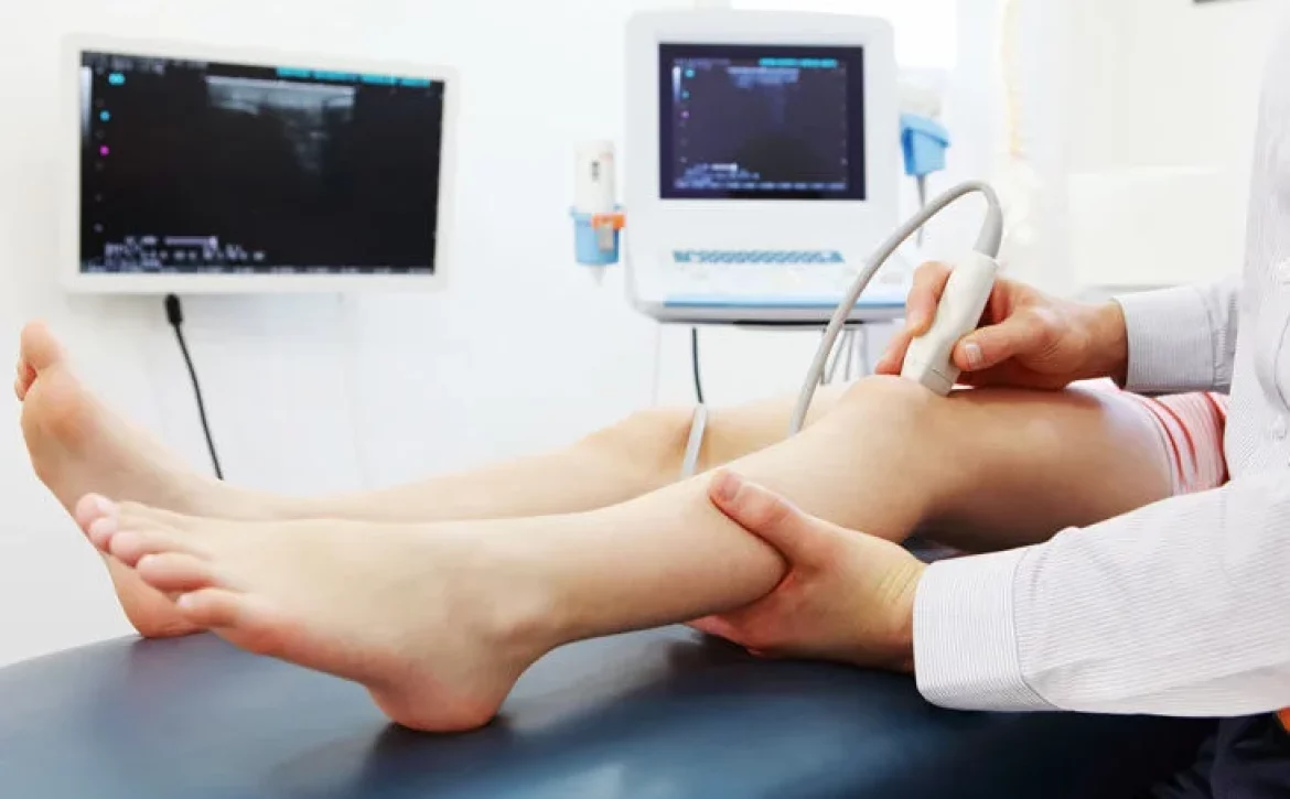
Foot ultrasounds are valuable diagnostic tools that provide detailed insights into the structures and health of the feet. This non-invasive imaging technique utilizes high-frequency sound waves to create real-time images of the internal structures, allowing healthcare professionals to assess injuries, conditions, and overall foot health. In this blog post, we will delve into the world of foot ultrasounds, exploring their uses, benefits, procedure, and what to expect during the examination.

Understanding Foot Ultrasound

Foot ultrasound, also known as sonography, uses sound waves that bounce off tissues to create images. It is commonly used to evaluate various conditions affecting the feet, including but not limited to:

  1. Soft Tissue Injuries: Foot ultrasounds can effectively visualize ligament, tendon, and muscle injuries, helping healthcare providers diagnose conditions like sprains, strains, and tears.

  2. Plantar Fasciitis: This common condition involves inflammation of the plantar fascia—a thick band of tissue running across the bottom of the foot. Ultrasound can aid in identifying the extent of inflammation and any associated tissue damage.

  3. Bursitis: Ultrasound can identify inflamed bursae (fluid-filled sacs that cushion bones and tendons) in the foot, helping in diagnosing conditions like retrocalcaneal bursitis.

  4. Nerve Issues: Conditions such as Morton’s neuroma, a thickening of tissue around a nerve between the toes, can be visualized using ultrasound.

  5. Arthritis: Ultrasound can help assess joint health, detect inflammation, and monitor the progression of arthritic changes in the foot.

  6. Fractures: While X-rays are more commonly used to visualize bone fractures, ultrasound can be complementary in assessing soft tissue damage and associated complications.

The Foot Ultrasound Procedure

  1. Preparation: Generally, foot ultrasounds do not require any special preparation. You might be asked to wear loose and comfortable clothing, or you may need to wear a gown during the procedure.

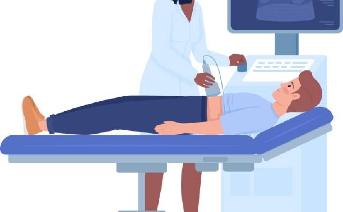
  2. Procedure: During the ultrasound, a technician (sonographer) will apply a clear gel to the area being examined. This gel helps the sound waves transmit effectively and prevents air gaps between the transducer (a handheld device) and your skin. The transducer is moved over the gel-coated area, emitting sound waves and capturing the returning echoes to create real-time images on a monitor.

  3. Duration: A foot ultrasound usually takes around 15 to 30 minutes, depending on the complexity of the examination.

  4. Painless and Non-Invasive: One of the major benefits of foot ultrasounds is that they are painless and non-invasive. There are no known risks associated with the procedure, making it safe for people of all ages.

What to Expect After the Ultrasound

After the foot ultrasound, you can immediately resume your regular activities. The clear gel will be wiped off, and you might experience some residual stickiness. The images captured during the procedure will be interpreted by a radiologist or healthcare provider, who will then share the results with you and discuss any further steps if necessary.

Closing Thoughts

Foot ultrasounds provide invaluable insights into the intricate structures of the feet, aiding in accurate diagnoses and effective treatment plans. Whether you’re experiencing foot pain, injuries, or other concerns, this non-invasive imaging technique can offer a clearer picture of what’s happening beneath the surface. If you’re experiencing foot-related issues, don’t hesitate to consult a medical professional who can determine if a foot ultrasound is the right diagnostic tool for you.

Prostate Ultrasound – What It Is and How It Is Done?

Prostate health is a crucial aspect of men’s overall well-being, and regular screenings play a vital role in detecting potential issues early on. One commonly used diagnostic tool is a prostate ultrasound. In this blog post, we will explore what a prostate ultrasound entails, its significance in prostate health, and how the procedure is performed. So, let’s dive in!

Can Ultrasound check prostate?

A prostate ultrasound is a non-invasive imaging technique that utilizes high-frequency sound waves to create detailed images of the prostate gland. It provides valuable insights into the size, shape, and condition of the prostate, aiding in the diagnosis of various prostate-related conditions.

The importance of an Ultrasound prostate scan

Prostate ultrasound is commonly used to evaluate and monitor conditions such as prostate cancer, benign prostatic hyperplasia (BPH), prostatitis, and other abnormalities. By visualizing the prostate gland, doctors can assess its structure, identify any abnormalities, and determine the appropriate course of action for further treatment or monitoring.

londonsono Testis Ultrasound Scan

How Prostate Ultrasound is Performed:

Preparation: Prior to the procedure, patients may be instructed to drink fluids and refrain from emptying their bladder to ensure better visibility during the ultrasound. It is advisable to follow the specific guidelines provided by the healthcare professional.

Positioning: The patient typically lies on their side or back, with their knees bent and pulled toward the chest. This position allows for easier access to the prostate gland.

Transrectal Ultrasound: A lubricated ultrasound probe, known as a transducer, is gently inserted into the rectum. The transducer emits sound waves that bounce off the prostate gland, creating real-time images on a monitor.

Image Capture: The ultrasound technician or radiologist moves the transducer within the rectum to capture images of different sections of the prostate gland. This process may take a few minutes to complete.

Post-Procedure: Once the images are obtained, the transducer is removed, and the procedure is complete. Patients can resume their normal activities immediately after the ultrasound.

Benefits and Considerations

Prostate ultrasound offers several advantages, including its non-invasive nature, real-time imaging capabilities, and the ability to visualize the prostate gland directly. However, it is important to note that prostate ultrasound alone may not provide a definitive diagnosis. In some cases, additional tests such as prostate biopsy or blood tests may be necessary to confirm or rule out certain conditions.

Prostate Health Check

Arranging an appointment for a Prostate Health Check

Prostate ultrasound is a valuable diagnostic tool in assessing prostate health and detecting potential abnormalities. By providing detailed images of the prostate gland, this procedure aids in the early detection and management of various prostate-related conditions. If you have concerns about your prostate health, consult with your healthcare provider, who can guide you on the appropriate screenings and examinations, including a prostate ultrasound if needed.

Remember, proactive monitoring and early detection are crucial in maintaining optimal prostate health, so prioritise regular check-ups and discussions with your healthcare professional. If you would prefer to arrange your ultrasound and health-checks yourself, you can do so directly with London Private Ultrasound, without the need for referral.

Simply contact us or visit our Prostate and Bladder Ultrasound Scan booking page to confirm the appointment date and time online.

Why do you need a full bladder for a pelvic ultrasound?

When you schedule a pelvic ultrasound, your healthcare provider may instruct you to arrive with a full bladder. You might wonder why a full bladder is necessary for this imaging procedure. In this blog post, we will explore the reasons behind the requirement of a full bladder for a pelvic ultrasound.

What is a pelvic ultrasound?

A pelvic ultrasound is a non-invasive imaging test that uses sound waves to create detailed images of the organs and structures within the pelvic region. It is commonly used to evaluate the uterus, ovaries, fallopian tubes, and bladder in women. In men, a pelvic ultrasound may be performed to examine the prostate gland and bladder. This diagnostic tool helps healthcare professionals assess the health and functionality of these organs, detect any abnormalities or conditions, and guide further treatment decisions.

Why is a full bladder necessary?

Improved Visualization: A full bladder acts as a “window” that helps enhance the visibility and clarity of the pelvic organs during the ultrasound. The filled bladder pushes the intestines and bowel loops away from the pelvis, creating a clear pathway for the ultrasound waves to travel through the pelvic region. This allows the sonographer or radiologist to obtain better images and capture more accurate details of the organs being examined.

Reference Point: The bladder serves as a reference point during a pelvic ultrasound. By having a full bladder, the healthcare professional can use it as a landmark to precisely locate and assess the position, size, and orientation of the pelvic structures. This aids in accurate measurements and evaluations.

Diagnostic Accuracy: Certain pelvic conditions and abnormalities can be better evaluated with a full bladder. For example, in women, a full bladder helps assess the position of the uterus, detect any uterine abnormalities, and evaluate the thickness of the uterine lining (endometrium). In men, a full bladder can help evaluate the size and shape of the prostate gland and detect any bladder-related issues.

Visit our service page: Bladder & Pelvic Ultrasound

Pelvic Ultrasound scan

How to prepare for a pelvic ultrasound

To ensure a successful pelvic ultrasound examination, it’s essential to follow the preparation instructions provided by your healthcare provider. Here are some general guidelines for preparing for a pelvic ultrasound:

Hydration: Drink plenty of water in the hours leading up to the ultrasound appointment to ensure your bladder is adequately filled. Your healthcare provider will provide specific instructions on how much water to drink and when to start drinking.

Timing: It’s important to time your fluid intake appropriately. Typically, you may be instructed to drink water about one hour before the scheduled appointment and avoid emptying your bladder until the ultrasound is complete.

Comfortable Attire: Wear comfortable and loose-fitting clothing that allows easy access to the pelvic area. You may be asked to change into a gown for the procedure.

Follow Instructions: Adhere to any additional instructions given by your healthcare provider, such as avoiding urination or emptying the bladder until after the ultrasound.

In conclusion, a full bladder is necessary for a pelvic ultrasound due to several reasons. It helps improve visualization, serves as a reference point for accurate measurements, and enhances diagnostic accuracy by aiding in the evaluation of pelvic organs and structures. By following the preparation guidelines provided by your healthcare provider, you can ensure a successful pelvic ultrasound examination and assist the healthcare team in obtaining accurate and valuable information about your pelvic health.


August 11, 2023 by Londonsono 0 Comments

Understanding Fatty Liver: Causes, Symptoms, and Management

Fatty liver, medically known as hepatic steatosis, is a common condition characterized by the accumulation of excess fat within the liver cells. This condition can have various causes and can range from mild to more severe forms. Fatty liver is becoming increasingly prevalent worldwide, and it’s important to understand its causes, symptoms, and management strategies.

Causes of Fatty Liver:

Non-Alcoholic Fatty Liver Disease (NAFLD): The most common cause of fatty liver is non-alcoholic fatty liver disease, which is often associated with obesity, insulin resistance, and metabolic syndrome. These conditions contribute to the accumulation of fat within the liver cells.

Alcoholic Fatty Liver Disease: Excessive alcohol consumption over an extended period can lead to alcoholic fatty liver disease. Alcohol is processed in the liver, and heavy drinking can overwhelm the liver’s ability to metabolize fat.

Medications and Toxins: Certain medications, such as corticosteroids and tamoxifen, as well as exposure to toxins, can lead to fatty liver.

Rapid Weight Loss: Sudden and significant weight loss, especially in cases of obesity, can cause the liver to release fat into the bloodstream, leading to fatty liver.

Symptoms of Fatty Liver:

In its early stages, fatty liver may not cause noticeable symptoms. However, as the condition progresses, you may experience:

Fatigue

Unexplained weight loss

Pain or discomfort in the upper right abdomen

Enlarged liver

Elevated liver enzymes in blood tests

It’s important to note that these symptoms can be indicative of other conditions as well. Therefore, if you’re experiencing any of these symptoms, it’s crucial to consult a medical professional for proper evaluation.

If you are unsure of the symptoms you are experiencing we would recommend an abdominal ultrasound, this will allow the assessment of other possible causes of symptoms in the abdomen and liver such as diagnosing gallstones, tumours, cysts and also liver function.

Diagnosis and Risk Factors:

Fatty liver can be diagnosed through imaging studies such as ultrasound, CT scans, and MRI. These tests help visualize the amount of fat accumulated in the liver. Your doctor may also recommend blood tests to assess liver function and rule out other potential causes.

Certain factors increase the risk of developing fatty liver:

Obesity or overweight

Type 2 diabetes

High blood pressure

High cholesterol levels

Sedentary lifestyle

Poor diet

Rapid weight loss

Management and Prevention:

Managing and preventing fatty liver involves lifestyle modifications and, in some cases, medical intervention:

Healthy Diet: Adopting a balanced diet rich in fruits, vegetables, whole grains, lean proteins, and healthy fats can help prevent and manage fatty liver. Avoiding high-calorie, sugary, and processed foods is crucial.

Regular Exercise: Engaging in regular physical activity helps improve insulin sensitivity, aids in weight loss, and reduces fat accumulation in the liver.

Weight Management: Maintaining a healthy weight is essential for preventing and managing fatty liver. Gradual, sustainable weight loss can significantly improve liver health.

Limit Alcohol Consumption: If you have alcoholic fatty liver disease, reducing or eliminating alcohol consumption is crucial for recovery.

Medical Supervision: In some cases, medical intervention may be required. Your doctor may prescribe medications to manage underlying conditions, such as diabetes or high cholesterol.

How do you know if you have a fatty Liver?

Fatty liver is a prevalent condition that can have serious implications for your liver health. Understanding its causes, symptoms, and management strategies is essential for preventing its progression and ensuring optimal liver function. If you suspect you have fatty liver or are at risk, consult a medical professional for proper evaluation and guidance.

Remember, a healthy lifestyle, including a balanced diet, regular exercise, and avoiding excessive alcohol consumption, plays a key role in maintaining your liver’s well-being. Prioritising your liver health today can lead to a healthier tomorrow.

We offer a range of scans suitable for those wanting a diagnosis on fatty liver disease and the option below allows you to select your appointment date and time for a Liver Fibroscan additionally we offer a Liver and Pancreas screening blood test or a liver ultrasound option.

August 10, 2023 by Londonsono 0 Comments

Kidney

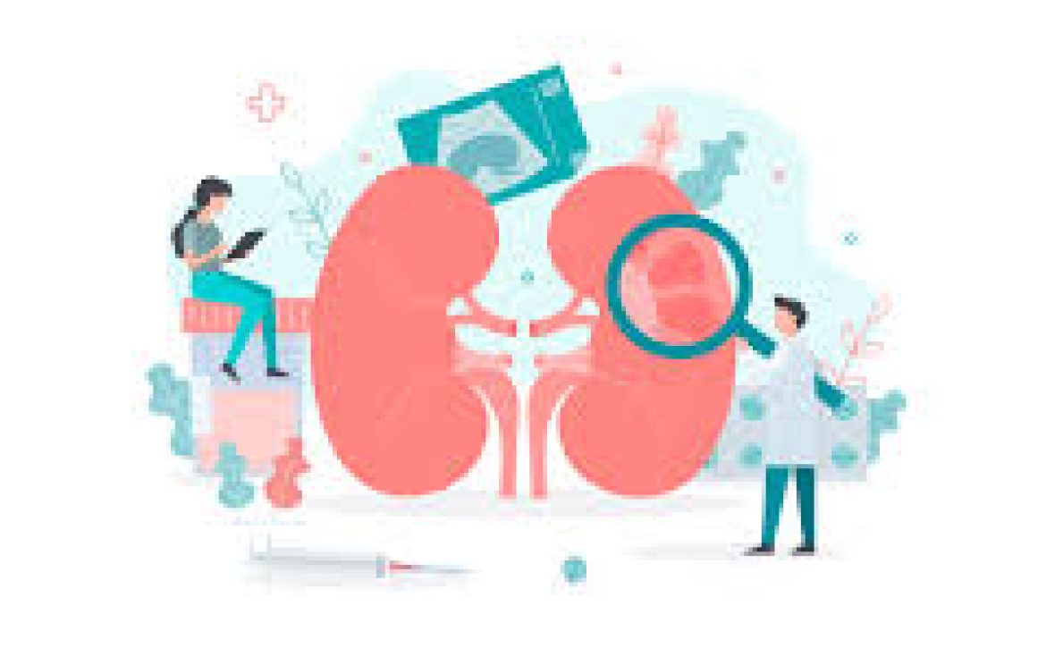
At our ultrasound clinic in London, we offer a comprehensive Kidney Cancer Package to support the early detection and management of kidney cancer. Our dedicated team of professionals utilizes advanced ultrasound technology to provide accurate assessments. Here’s how we can assist you:

Blood and urine tests: Our clinic perform specialized blood and urine tests to detect any abnormalities that may indicate kidney cancer. These tests help assess levels of red blood cells, white blood cells, and various enzymes that can be indicative of the disease.

Kidney and Urinary Tract ultrasound: Our advanced ultrasound scans specifically focus on the kidneys and urinary tract. This non-invasive procedure allows us to detect the presence of tumors or nodules, determine their size, location, and assess whether they have spread to other parts of the body.

GP Consultation: We offer consultations with experienced general practitioners who specialize in kidney health. They will assess your symptoms, review your test results, and provide expert guidance and recommendations tailored to your specific needs.

Early detection and treatment are crucial for improving outcomes in kidney cancer. Our Kidney Cancer Package combines state-of-the-art ultrasound technology with expert medical care to ensure the best possible diagnosis and management.

If you have any concerns about kidney health or if you are at risk of kidney cancer, don’t hesitate to contact us and schedule an appointment. Your well-being is our priority, and we are here to support you every step of the way.

August 10, 2023 by Londonsono 0 Comments

Heart Health

Central London Heart Clinic

At our ultrasound scan clinic in London, we are dedicated to promoting heart health, and we offer a comprehensive Heart Health Package to ensure your well-being. The package includes an Echocardiogram (ECHO), a non-invasive test that utilizes ultrasound technology to generate detailed images of your heart. Our skilled Cardiac Physiologist will conduct the test, and the report will be reviewed by our Consultant Cardiologist Clinical Lead.

The Echocardiogram is a valuable tool for early detection and diagnosis of various heart conditions. It can identify heart disease, detect abnormalities, and guide appropriate treatment plans. If you experience symptoms such as breathlessness, persistent ankle swelling, a chronic cough, rapid heart rate, or unexplained weight loss, an Echocardiogram can provide crucial insights.

The scan allows us to evaluate the function of the left and right ventricles, detect heart failure, assess for valvular heart disease, cardiomyopathy, pericardial disease, and aortic dissection. It is also an effective diagnostic tool for conditions like coronary artery disease and cardiac arrhythmia, and it can help investigate heart-related symptoms such as chest pain and dizziness.

To complement the Echocardiogram, we also offer GP consultations with experienced practitioners specialising in heart health. They will assess your symptoms, review test results, and provide expert guidance tailored to your specific needs. Early detection and management of heart problems are crucial for optimal health.

If you have any concerns about your heart health, don’t hesitate to contact us and schedule an appointment. Your well-being is our priority, and we are here to support you every step of the way.

Private Echocardiogram Clinic

The Echocardiogram is a valuable tool for early detection and diagnosis of various heart conditions. It can identify heart disease, detect abnormalities, and guide appropriate treatment plans. If you experience symptoms such as breathlessness, persistent ankle swelling, a chronic cough, rapid heart rate, or unexplained weight loss, an Echocardiogram can provide crucial insights.

The scan allows us to evaluate the function of the left and right ventricles, detect heart failure, assess for valvular heart disease, cardiomyopathy, pericardial disease, and aortic dissection. It is also an effective diagnostic tool for conditions like coronary artery disease and cardiac arrhythmia, and it can help investigate heart-related symptoms such as chest pain and dizziness.

To complement the Echocardiogram, we also offer GP consultations with experienced practitioners specialising in heart health. They will assess your symptoms, review test results, and provide expert guidance tailored to your specific needs. Early detection and management of heart problems are crucial for optimal health.


If you have any concerns about your heart health, don’t hesitate to contact us and schedule an appointment. Your well-being is our priority, and we are here to support you every step of the way.

Cost of seeing a cardiologist privately uk

If you have any concerns about your heart health, don’t hesitate to contact us and schedule an appointment. Your well-being is our priority, and we are here to support you every step of the way. We have a range of heart services available to book privately and often, via online booking you can book a same day or next day clinic appointment, without any need for GP referral.


We offer a range of Cardiologist appointments and also some specific services for those of you concerned about cholesterol or diabetes and also Full Body scans and total body health checks – so you can take advantage of one-stop clinics that provide the exact combinations of blood and body tests, ultrasound imagery, reports and consultations.

Prices range from £80 for an ECG to around £200 for most standard heart clinics, we also provide a platinum cholesterol health service which includes a full suite of ultrasound and heart diagnostic tests, reports and consultation.

Ovarian Cancer Awareness

Ovarian Cancer Awareness Month is in March. The purpose of this month is to raise awareness about ovarian cancer, promote early detection, and support those who have been affected by the disease.

Ovarian cancer is a group of different cancers that originates in the ovaries. Ovaries are two in number and part of female reproductive organs which produce eggs. Ovarian cancer in its early stages has no particular signs and it can be difficult to detect in its early stages, therefore it is why raising awareness about the symptoms and risk factors is so important.

During Ovarian Cancer Awareness Month, people are encouraged to learn about the symptoms and the risk factors of ovarian cancer.

In addition to raising awareness, Ovarian Cancer Awareness Month is also a time to support those who have been affected by the disease. This may involve participating in fundraising events, donating to ovarian cancer research organizations, or simply offering emotional support to loved ones who have been impacted by the disease.

Overall, Ovarian Cancer Awareness Month is an important opportunity to educate people about the risks and symptoms of ovarian cancer and to advocate for increased research and funding to improve early detection and treatment options for those affected by the disease.

Signs of ovarian cancer

Ovarian cancer may not cause any specific symptoms in the early stages. However, as cancer grows or spreads, it can cause a range of symptoms. Here are some common signs and symptoms of ovarian cancer:

  1. Abdominal or pelvic pain.
  2. Bloating or feeling full quickly.
  3. Changes in bowel habits: Constipation or diarrhea.
  4. Frequent urination.
  5. Loss of appetite and weight.
  6. Fatigue.
  7. Abnormal vaginal bleeding.

It’s important to note that these symptoms can be caused by many conditions other than ovarian cancer. However, if you experience any of these symptoms for an extended period of time, or if they are accompanied by other signs, you should see a doctor for further evaluation. Early detection is key to the successful treatment of ovarian cancer.

Factors that can increase the risk of ovarian cancer

There are several factors that can increase a woman’s risk of developing ovarian cancer. Some of the most common risk factors include:

  1. Age: The risk of developing ovarian cancer increases as a woman gets older. Most cases of ovarian cancer occur in women over the age of 50.
  2. Family history: Women with a family history of ovarian cancer are at an increased risk of developing the disease. This risk is higher if a first-degree relative, such as a mother, sister, or daughter, has been diagnosed with ovarian cancer.
  3. Inherited genetic mutations: Some inherited genetic mutations, such as those in the BRCA1 and BRCA2 genes, can increase a woman’s risk of developing ovarian cancer.
  4. Reproductive history: Women who have never been pregnant or who had their first pregnancy after age 35 are at a slightly higher risk of ovarian cancer. Women who have had multiple pregnancies and/or used birth control pills for an extended period of time are at a lower risk.
  5. Hormone replacement therapy (HRT): Some studies suggest that long-term use of HRT, particularly estrogen-only HRT, may increase a woman’s risk of developing ovarian cancer.
  6. Endometriosis: Women with endometriosis, a condition in which tissue that normally lines the inside of the uterus grows outside of it, are at an increased risk of developing ovarian cancer.

It’s important to note that having one or more of these risk factors does not mean that a woman will develop ovarian cancer. However, if you have one or more of the above risk factors, you should discuss your risk and appropriate screening options with your doctor.

Detecting ovarian cancer

Ultrasound can help detect ovarian cancer. Transvaginal ultrasound is one of the primary imaging tests used to diagnose ovarian cancer. It uses high-frequency sound waves to create images of the ovaries and surrounding tissues.

During a transvaginal ultrasound, a small wandlike instrument or probe is inserted into the vagina, which allows for a closer view of the ovaries and surrounding structures. The procedure is painless and takes only a few minutes to complete.

Transvaginal ultrasound can help detect ovarian tumors or masses, as well as changes in the size or shape of the ovaries, which might indicate the presence of cancer. If a tumor is detected, the ultrasound can also help determine its size, location, and whether it is a solid or fluid-filled, cystic.

However, it’s important to note that transvaginal ultrasound is not a definitive test for diagnosing ovarian cancer. If an abnormality is detected on the ultrasound, further testing, such as a blood test or biopsy, may be needed to confirm whether the mass is cancerous.

You can book your pelvic scan (two parts: transabdominal and transvaginal examination) with London Private Ultrasound Clinic located on Welbeck Street. Our medical ultrasound specialists are fully qualified with more than 10 years of experience. We offer same-day appointments with evening and weekend availability. You can book your appointment very easily either through our website or through a phone call on 020 7101 3377.

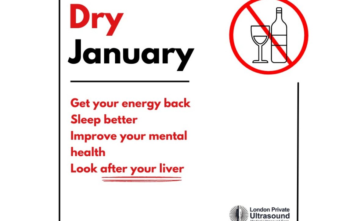
Celebrating ten years of Dry January

Committing to Dry January

January is when you should start fulfilling those New Year’s Resolutions. And maybe one of them was to consume less alcohol. We’re with you on this one. You might wonder about the rules of this challenge. It’s pretty simple—no alcohol from New Year’s Day or at least the 2nd of January until the 1st of February.

Not only will it be a fantastic way to regain energy, sleep better, and save money, but it will also help you be kind to your liver.


How many Alcohol units per week?

It is usually recommended not to drink more than 14 units of alcohol per week. Even though your liver can handle small amounts of alcohol, it can still affect it in many ways. It might be possible that you’re damaging your liver, and you don’t even know about it. If you feel any pain on the right side of your abdomen when drinking alcohol, you should have an abdominal ultrasound scan.

It is also best to allow yourself at least 1-2 days alcohol free per week.

Look after your liver.

The best way to check on your liver is to have an ultrasound scan which can rule out any abnormality, fatty liver disease or other alcohol-related liver diseases. If you don’t want to wait 6+ weeks for your hospital appointment, book a private abdominal ultrasound scan in our Private Ultrasound Clinic in Welbeck Street, just a few mins walk from Bond Street tube station. We offer many packages to help you assess your liver health including a liver scan, abdominal scan and blood tests and can provide you with a verbal summary during the appointment as well as a written report. We can provide you with a KPA number, which will allow you to determine whether your liver health has improved or declined upon your second visit. We would recommend the liver elastography for a KPA assessment of your liver.

We offer same-day, weekend, and evening appointments for an affordable price.

What is a kPa score?

The health of your liver and the elasticity of the liver is typically measures on a scale of 1-14+ with kilopascals (kPa).

The measurement score allows us to determine the appropriate next steps and generally the scores will be provided to assess the following states – little or no scarring, moderate scarring, intense scarring and irreversible scarring. It is important information to know as there is much you can do to heal the liver and as such, one reason why we encourage all our patients to observe Dry January and take all the lifestyle steps to improve your general health and reduce toxicities, particularly those of the liver.

Your liver plays a pivotal role in your body’s detoxification processes and overall health. Excessive alcohol consumption can put significant strain on this vital organ, potentially leading to liver diseases such as fatty liver disease, hepatitis, or even cirrhosis. Our advanced ultrasound services can provide crucial insights into your liver’s health, providing you with the optimal chance for a full recovery or control of your liver health.

Top tips for succeeding your Dry January Challenge

During Dry January, embracing alcohol-free alternatives can be a refreshing and health-conscious choice. Swap your usual alcoholic beverages for a variety of non-alcoholic options, such as alcohol-free beers, mocktails, or herbal teas. 

Experiment with different flavours to find what satisfies your taste buds and invest in some cocktail kits to get creative and focus the mind on something more than just the drinking of it!

Meeting friends, can often mean drinking alcohol and if you feel you may crumble in this situation perhaps try arranging less tempting activities like coffee catch-ups, hiking, or attending cultural events. Opt for activities that don’t revolve around alcohol to create a supportive environment. 

The other vital element is taking up exercise, this can also be a daunting prospect if it has been years since you set foot in the gym or met-up for your weekly game! Enhancing blood flow and circulation, incorporating regular exercise into your routine, focusing on activities like brisk walking, cycling, or yoga all help your liver to recover. 

Stay well-hydrated by drinking plenty of water throughout the day, aiding in the detoxification process. Additionally, prioritise a balanced diet rich in antioxidants from fruits, vegetables, and whole grains. Consider incorporating foods like ginger and garlic known for their circulation-boosting properties. Adequate sleep is crucial for overall health, so ensure you get quality rest each night. 

Finally, take time for relaxation through practices like deep breathing or meditation to manage stress, supporting both physical and mental well-being during your alcohol-free month. Perhaps induce a more relaxing environment by indulging in a little shopping for comforts, candles and planning your holidays!

Drinks making Alcohol free easy

  1. Non-Alcoholic Beers: Many breweries now produce alcohol-free versions of their beers. Brands like Heineken 0.0, Budweiser Zero, and Clausthaler Original offer an alcohol-free option with the same beer flavor. For those of you more on the craft beer scene, the options will not disappoint – try this link to the independent and their top selections of low and no-alcohol beers.

  2. Non-Alcoholic Wines: There are various alcohol-free wines available that mimic the taste and complexity of traditional wines. Look for brands such as Ariel Vineyards or Torres Natureo, most supermarkets offer a particularly good selection to choose from – you may even find a alcohol free English sparkling wine to celebrate in style through Dry January.

  3. Mocktails and Mixers: Some companies specialize in crafting alcohol-free versions of classic cocktails. Brands like Seedlip offer a range of non-alcoholic spirits that can be used to create sophisticated mocktails. Additionally, there are non-alcoholic versions of popular mixers like tonic water or ginger beer.

  4. Non-Alcoholic Spirits: Beyond traditional mocktails, some brands focus on creating alcohol-free alternatives to spirits like gin, vodka, or rum. Examples include Seedlip Grove 42 (citrus-focused) or Ritual Zero Proof (gin alternative).

  5. Soft Drinks and Infused Waters: Explore a variety of artisanal sodas, flavored sparkling water, and infused waters. Brands like Fentimans, Fever-Tree, or LaCroix offer unique and refreshing options.

Pregnancy Ultrasound Scan

Antenatal Scans and Screenings:
Pregnancy is a very exciting time, doesn’t matter if this is your first or not. It is especially important to look after yourself and your unborn baby by getting familiar with your antenatal care or “Pregnancy Care”. “Antenatal or Pregnancy Care” is a package of healthcare and support you should have while you are pregnant. This care package is in place to make sure you and your unborn baby are safe and well throughout your pregnancy journey. Regular Antenatal care is important to:

  1. Keep an eye on how your baby is growing
  2. Pick up conditions that might not have any early symptoms to notice but routine checks can notice them even you feel perfectly fine.
  3. Check the health of your baby through blood tests and ultrasound scans (find more information about ultrasound scans and blood tests below).

Ultrasound scan: Ultrasound scan is the safest way to look at your baby in your womb. An ultrasound scan can tell the approximate date your baby is due, confirm whether you are having more than one baby, and notice some possible pregnancy complications. We know that seeing your baby for the first time can feel like an exciting event and that you may feel anxious too. Therefore, you can bring your partner, a friend, or a family member along with you to share it.

How does the ultrasound scan work? Ultrasound is a safe and non-invasive imaging technique using high-frequency sound waves to produce black and white or color images of your baby, which has been around for half a century with no known harm to the unborn child. All medical care pregnancy relies heavily on ultrasound findings during pregnancy. Pregnancy is divided into 3 trimesters each lasting around 12 weeks which makes the length of the pregnancy 38-42 weeks approximately. During the 3 trimesters of pregnancy different ultrasound imaging and techniques are used to help in the management of regular monthly care and medical or surgical intervention if needed.


First Trimester: The duration of the first trimester begins from the first day of your last period up to week 12 of your pregnancy. From the first day of your last period up to week 12 of your pregnancy is the first trimester. During this time, the fertilized egg and sperm, the new baby cells, implant themselves inside the lining of the womb(endometrium). Sometimes this does not happen, and the pregnancy starts to grow outside the womb and is called an ectopic pregnancy, which is a medical/surgical emergency. Sometimes there might be hidden bleeding inside with the possibility of miscarriage or the tissue of the placenta becomes abnormal which is called molar tissue both of which need medical intervention.

In the early stages of the first trimester between 5-7 weeks, ultrasound has an invaluable diagnostic power. Two tools of external scanning (Transabdominal with a full bladder) and internal (Transvaginal with empty bladder) ultrasound is used to diagnose the followings:

  • 1) Determination of the site of the pregnancy sac whether it the correctly placed in the womb or abnormally and ectopically outside the womb.
  • 2) Determination of the number of pregnancy sacs and the number of babies present in single or multiple pregnancies.
  • 3) Determination of viability of the baby by visualization of the heartbeat. This usually starts around 5-6 weeks of gestation. A rescan is always offered if a heartbeat cannot be seen in very early gestations to recheck the viability a few days later.
  • 4) Determination of the possibility of complications such as bleeding, possibility of miscarriage, or abnormality of the tissue of the placenta such as molar pregnancy.
  • 5) Determination of the health of ovaries and the corners of your pelvis which is called adnexa.
  • 6) During 5-7weeks baby appears as a bean-shaped structure with no arms and legs and the presence of a heartbeat is considered a baby’s health.


Later, at around 10 weeks, the baby’s arms and legs and top of the head can be seen and internal structures start to show themselves, and the spine and front of the abdomen can also be checked.
From around 10 and 4 days to 12 weeks the baby is big enough to be checked for possible chromosomal abnormalities. The two most common tests available are based on ultrasound scan and a blood test from the mother’s arm are NIPT and NT screening.


Second trimester: During the second trimester of the pregnancy from 12 weeks to 24 weeks, the baby’s structures are fully formed and can be checked by ultrasound. Gradually from 16 weeks onwards, the baby’s gender (Sex) can be identified. During this period, the baby’s health and anatomy are checked. Development of the brain and heart and all internal organs are checked. Further, along with from 18-22 weeks with an anomaly scan which is a very detailed scan, the baby’s growth, health, heart, spine, abdomen, and kidney structures can be checked in detail. Fingers and toes are counted for. The placenta position is checked to be high in relation to the cervix to allow safe passage for a natural delivery. Blood flow from the placenta to the baby is also checked by color Doppler imaging. With an anomaly scan completed, usually, no other scan is offered to mothers under NHS. However, with a reassurance scan that looks at any concerns, you might have during the 2nd and 3rd trimesters you can have a detailed scan checking your concerns and have a piece of mind.


When can I tell if I am having a boy or girl? At your 16-40 weeks of pregnancy, the sonographer may be able to see if your baby is a boy or a girl. Tell them if you would like to know – and tell them too if you don’t want to find out until the birth.


Third trimester: From 24 weeks onwards your fully formed baby starts to put on weight and grow. Growth scans are set to examine babies who on routine monthly maternity examinations by GP or midwife are either suspiciously too big or too small on maternal fundal height measurements. Some mothers can develop gestational diabetes or have high blood pressure or any other medical history or complications which might affect the growth of the baby. You might be just curious about the weight of your baby or anxious over the function of the placenta. Growth scans are designed to check the baby’s estimated weight by measuring the baby’s head circumference, abdomen circumference, and the baby’s length of the thigh bone measurements. The blood flow from the placenta to the baby is checked to ensure placental health, amniotic fluid is measured and the baby’s internal organs such as heart, lungs, diaphragm, kidneys, stomach, and bladder are also checked.
Between 24-32 weeks, if you are interested to see your baby in real-time imaging or still images in color imaging you can book a 3/4D scan. You may be lucky to see him/her sucking their thumb, have a yawn, stretch, smile, or frown and see your baby’s face in real-time and further bond with your little bundle of joy.

Presentation scan: Some pregnancies might need a much later scan as there might be a chance that the baby’s head is not down and maybe breech or transverse. A presentation scan is performed from 36-37 onwards and it not only checks the position of the head and gives information about the presentation and a safe passage for delivery but also checks all information regarding the baby’s growth and wellbeing, blood flow from the placenta and amniotic fluid volume is checked.


Cervical length scan: Some women have a history of late 2nd and 3rd-trimester miscarriages in their previous pregnancies, some are worried about the possibility of a shortened cervix, and some have a history of surgery on the cervix, and all are worried to know if their cervix is long enough, closed (competent) and able to keep the pregnancy for its duration. Transvaginal Cervical length measurement is a safe non-invasive ultrasound method to show the length and competency of the cervix. It can be performed from 16 weeks gestation. It also can help to figure out if the placenta is on the cervix (placenta previa) or a loop of the umbilical cord is trapped underneath the baby’s head or leg/feet near the cervix called cord presentation. If a transvaginal scan is prohibited or undesired by the patient trans labial scan (Scanning between the mother’s leg over the genital area) is the second-best choice to perform an examination to get accurate measurements.


Screening and diagnostic tests: Screening tests will let you know whether your baby has a high chance of particular conditions such as Down’s syndrome, Edwards syndrome, and Patau syndrome. At London Private Ultrasound we can provide you with the most completed package of screening tests as follows:


NT and NIPT TestsThese two tests are non-invasive screening tests that check three major chromosomal abnormalities in early pregnancy. Trisomy 21 or Downs syndrome, Trisomy 18 or Edwards syndrome, and Trisomy 13 or Patau syndrome.


NIPT (Non-invasive prenatal test)This test is carried out as early as 10 weeks and 4 days. A dating or an early ultrasound scan is performed, and the gestational age is established. The baby’s body is checked thoroughly, and structures are identified to be normal. Then Blood is drawn from the mother’s arm. The blood is sent to the lab and is analyzed for the baby and placental cells floating in the mother’s bloodstream. With NIPT the baby’s gender(sex) and health of sex chromosomes can be checked also. Once all is established a comprehensive report is issued which gives the risk factor of the trisomies mentioned above. NIPT overall is 99% accurate.


NT screening Test: This test is performed from 11 weeks to 13 weeks and 6 days. An early or dating scan is performed, and the gestational age is established. The baby’s body is checked thoroughly, and structures are identified to be normal. An advanced practitioner with relevant qualifications measures the thickness of the accumulation of fluid between two layers of the skin at the back of the baby’s neck. This is called nuchal translucency or NT. All babies have some fluid at the back of their neck at this stage of the pregnancy, however, babies who have thickened layers tend to have a higher risk of chromosomal abnormalities or heart, lymphatic, or kidney disease. So just having an increased thickness does not necessarily mean that the baby has chromosomal abnormality (the NT test on its own is only 70% accurate). By 14 weeks gestation, this fluid gradually disappears and cannot be measured accurately. Then Blood is drawn from the mother’s arm. The blood is sent to the lab and is analyzed. Blood test results are based on pregnancy hormone, or the placenta hormone called BHCG, and a protein produced by the placenta called Papp-A, which are then combined with ultrasound NT measurement, and then the risk is given as a comprehensive result based on these findings and the baby’s gestation. This combined test is about 80-90% accurate.

 

Thyroid Ultrasound Scan

Ultrasound is the imaging modality of choice when evaluating the thyroid. It is a safe, painless, and non-invasive procedure. The procedure uses high-frequency sound waves to produce images of the thyroid and other organs inside your body. Ultrasound provides excellent anatomic detail and plays a vital role in current clinical practice; with accurate diagnosis ultrasound significantly improves the management of numerous medical conditions. The thyroid is a gland situated at the front of the neck. It consists of two lobes that lay on either side of the windpipe (trachea) and are connected by a strip of thyroid tissue called the isthmus. The thyroid gland is an endocrine gland that produces and releases hormones into the bloodstream.

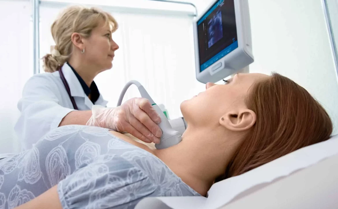
What happens during my scan? A thyroid ultrasound scan doesn’t need preparation. You just have to expose your neck. You will be examined in a semi-sitting position with your chin upwards. A high-frequency linear transducer array will be used to provide a high-resolution image. Greyscale and color doppler ultrasound are used to evaluate a thyroid lesion. The size, form, borders, echogenicity, contents, location, and vascular pattern of the whole thyroid gland will be assessed. In some cases, fine needle aspiration or biopsy might be recommended when further investigations are needed.


Can ultrasound detect any nodules in my thyroid? Ultrasound has become an essential diagnostic technique in the examination of thyroid nodules. It is highly sensitive in detecting nodules, the sonographic features, and whether they are benign nodules or if they have malignant characteristics that require further investigation such as a biopsy or FNA. Fine needle aspiration (FNA) or Biopsy can help to improve the accuracy of diagnosis.


What is a thyroid nodule? Thyroid nodules are categorized according to the “U” classification by the British thyroid association. Under this classification, Nodules are classified into categories (U1-U5) based on features including echogenicity, margins, internal echo pattern, calcification, and vascularity. (U1) indicates a normal thyroid. A benign thyroid nodule is indicated by (U2). A U3 nodule is indeterminate/equivocal. (U4) indicates a suspicious nodule and (U5) indicates malignancy. U3-U5 nodules require FNA or Biopsy to be performed.


What else can be detected in my thyroid scan? Thyroid ultrasound is also used to evaluate diffuse changes in thyroid parenchyma. Hashimoto thyroiditis and Graves’ disease are common disorders that present diffuse thyroid enlargement. Hashimoto thyroiditis is an autoimmune disease where the immune system attacks the thyroid gland therefore the thyroid is unable to produce enough hormones. Graves’ disease is another autoimmune disorder that causes the thyroid to become overactive and produce excess thyroid hormones thus several body functions work faster than usual.

>> Click on the link for online booking in the fastest time