27 Welbeck Street, London W1G 8EN
Tel: 020 7101 3377
August 12, 2023 by Londonsono 0 Comments

Navigating the Breast Ultrasound Procedure: A Comprehensive Guide

At London Private Ultrasound, we prioritize your health and well-being, offering advanced imaging services to ensure accurate diagnoses and personalized care. If you’ve been scheduled for a breast ultrasound, you may have questions about the procedure and what to expect. In this article, we’ll provide you with a comprehensive overview of the breast ultrasound procedure, from preparation to post-procedure care.

Understanding Breast Ultrasound: A Non-Invasive Imaging Technique

Breast ultrasound is a safe and non-invasive imaging technique that uses sound waves to create detailed images of the breast tissue. It is particularly effective in evaluating breast lumps, changes, or abnormalities detected during physical exams, mammograms, or self-examinations. Ultrasound allows healthcare professionals to visualize the internal structures of the breast, providing valuable insights for diagnosis and treatment.


Preparation: What to Do Before Your Breast Ultrasound

Preparing for a breast ultrasound is straightforward and generally requires minimal steps. Here’s what you can do to ensure a smooth experience:

Wear Comfortable Clothing: You’ll be provided with a gown to wear during the procedure, so opt for comfortable clothing that can be easily removed.

Avoid Lotions and Creams: To ensure the ultrasound transducer makes direct contact with the skin, refrain from applying lotions, creams, or powders to your breast area on the day of the procedure.

Share Relevant Information: Inform the ultrasound technologist about any breast symptoms, pain, or concerns you may have. This information helps ensure a thorough and accurate examination.

The Breast Ultrasound Procedure: What to Expect

The breast ultrasound procedure is a straightforward and painless process. Here’s a step-by-step guide to what you can expect during your visit to our clinic:

Welcome and Check-In: Upon arrival, you’ll be warmly greeted by our staff. They will guide you through the check-in process, ensuring that all necessary paperwork is completed.

Preparation: You’ll be taken to a private examination room where you can change into a gown. A friendly ultrasound technologist will explain the procedure, answer any questions you may have, and address any concerns.

Positioning: You’ll be asked to lie on an examination table. The technologist will help you position yourself comfortably, ensuring optimal access to the breast area.

Gel Application: A water-based gel is applied to the breast area. This gel facilitates the transmission of sound waves and ensures clear images are captured during the procedure.

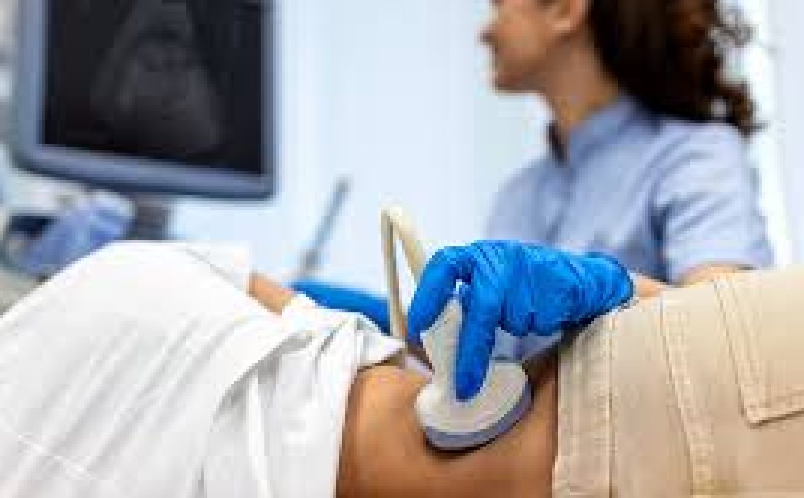
Transducer Placement: The ultrasound technologist will use a handheld device called a transducer. The transducer emits high-frequency sound waves that bounce off the breast tissue and create real-time images on a monitor.

Image Capture: The technologist will gently move the transducer over the breast area, capturing images from various angles. You may hear faint sounds as the sound waves are emitted and received.

Image Analysis: As images are captured, the technologist will analyze them to ensure comprehensive visualization of the breast tissue. Different angles and views may be explored to thoroughly assess any areas of concern.

Duration: The procedure is typically completed within 15 to 30 minutes, depending on the specifics of the examination.

After the Procedure: Post-Examination Steps

Following the breast ultrasound procedure, you can expect the following steps:

Gel Removal: The water-based gel applied to your breast area will be gently wiped off.

Results Discussion: In some cases, the ultrasound technologist may discuss initial findings with you. However, a comprehensive analysis of the images will be performed by a radiologist.

Radiologist’s Interpretation: The captured images will be reviewed and interpreted by a specialized radiologist. This expert analysis ensures accurate diagnosis and personalized recommendations.

Follow-Up Steps: Based on the radiologist’s interpretation, your healthcare provider will discuss the results with you and provide guidance on any necessary next steps.


Advantages of Breast Ultrasound:

Breast ultrasound offers several advantages that contribute to its importance in breast health:

Non-Invasive: Ultrasound is non-invasive and does not involve radiation exposure, making it a safe choice for breast imaging.

Distinguishing Characteristics: Ultrasound can differentiate between solid masses and fluid-filled cysts, aiding in the characterization of breast abnormalities.

Guidance for Biopsies: Ultrasound can guide the placement of a needle for biopsy, ensuring precise sampling of tissue for further analysis.

No Pain or Discomfort: The breast ultrasound procedure is painless and generally well-tolerated.

The breast ultrasound procedure is a crucial tool in maintaining optimal breast health. It provides valuable insights into breast tissue, aiding in the early detection and diagnosis of potential issues. At our ultrasound clinic, we are committed to providing you with a comfortable and informative experience during your breast ultrasound.

If you have questions about the breast ultrasound procedure, wish to schedule an appointment, or seek guidance on your breast health journey, our experienced team is here to support you every step of the way. Your well-being is our priority, and we are dedicated to empowering you with the knowledge and tools needed for optimal breast care.

August 12, 2023 by Londonsono 0 Comments

What is Breast implants, Different Types of Breast Implants and Importance of regular check up using Ultrasound scan

Breast implants are medical devices that are placed inside the breast tissue or chest muscles to enhance breast size or restore breast volume after surgery. They are often used for cosmetic reasons, such as breast augmentation, as well as for reconstructive purposes after mastectomy or other medical procedures.

Types of Breast Implants:

Silicone Implants: These implants are filled with silicone gel, which closely resembles the feel of natural breast tissue. They are known for their natural appearance and texture.

Saline Implants: Saline implants are filled with sterile saltwater solution. They are inserted into the breast empty and then filled with the saline solution. They can be adjusted for size during surgery.

Structured Saline Implants: These implants have an internal structure that controls the movement of the saline solution, giving them a more natural feel.

Gummy Bear Implants (Form-Stable Implants): These implants are filled with a thicker silicone gel that retains its shape even if the implant shell is damaged. They are known for maintaining their form over time.

Reasons for Getting Breast Implants:

Breast Augmentation: Many individuals opt for breast implants to enhance the size and shape of their breasts for cosmetic reasons.

Breast Reconstruction: Breast implants can be used for reconstructive purposes after a mastectomy or other breast surgeries.

Asymmetry Correction: Implants can help correct breast size or shape discrepancies between the two breasts.

The Surgical Procedure:

The procedure for getting breast implants typically involves the following steps:

Consultation: You’ll have a consultation with a plastic surgeon to discuss your goals, preferences, and any medical considerations.

Incision: The surgeon makes an incision in a discreet location, such as under the breast, around the areola, or in the armpit.

Implant Placement: The implant is inserted either behind the breast tissue (subglandular) or under the chest muscle (submuscular).

Closing the Incision: The incision is closed with sutures or surgical tape.

Recovery: Recovery time varies, but you may need to avoid strenuous activities for a few weeks and follow your surgeon’s post-operative care instructions.

Risks and Considerations:

While breast implants can yield satisfying results, it’s important to be aware of potential risks and considerations:

Rupture or Leakage: Implants can rupture or leak over time, leading to changes in breast shape or texture.

Capsular Contracture: Scar tissue may form around the implant, causing the breast to feel firm or misshapen.

Infection: Infections can occur after surgery, necessitating removal of the implant.

Implant Displacement: Implants may shift or move from their original position.

Changes Over Time: Implants are not lifetime devices and may need to be replaced after a certain period.

Regular Monitoring:

It’s important to have regular follow-up appointments with your surgeon and undergo recommended imaging, such as mammograms or ultrasounds, to monitor the condition of your breast implants.

Considerations for Ultrasound Clinics:

For ultrasound clinics, breast imaging services can play a crucial role in monitoring breast implants. Ultrasound imaging can help evaluate the integrity of the implant, detect leaks or ruptures, and assess any changes in breast tissue. It’s especially useful for individuals with silicone implants, as ultrasound can effectively visualize the implant and surrounding tissues.

Breast implants are medical devices that can enhance breast size, restore volume, and aid in breast reconstruction. They are used for various reasons, including cosmetic enhancement and medical purposes. While breast implants can yield positive outcomes, it’s important to thoroughly research and discuss the procedure with a qualified plastic surgeon. Regular monitoring and follow-up appointments, including ultrasound imaging, are essential for maintaining optimal breast health and the longevity of the implants.

As a trusted ultrasound clinic (London Private Ultrasound), we are dedicated to providing comprehensive imaging services, including ultrasound checks, to support individuals with breast implants. If you have any questions or concerns about breast implant monitoring or related imaging, our experienced team is here to assist you and ensure your breast health journey is well-guided and informed.

August 12, 2023 by Londonsono 0 Comments

Understanding Breast Cancer Symptoms: Early Detection and Awareness

Breast cancer is a significant health concern that affects both women and men, although less commonly, men. Early detection is key to successful treatment and improved outcomes. As a leading ultrasound clinic, we are dedicated to raising awareness about breast cancer symptoms to empower individuals to prioritize their breast health. In this article, we will explore the common symptoms of breast cancer and emphasize the importance of regular screenings.

Common Breast Cancer Symptoms:

Lump in the Breast or Underarm:

The most common symptom of breast cancer is the presence of a lump or mass in the breast or underarm. While not all lumps are cancerous, any new lump or change in the breast tissue should be promptly evaluated by a healthcare professional. It’s important to note that some breast cancers can be felt but not seen on imaging.


Changes in Breast Size, Shape, or Appearance:

Breast cancer may cause visible changes in the breast, such as swelling, dimpling, or distortion of the breast contour. These changes can occur due to the growth of a tumor affecting the surrounding tissue.


Unexplained Nipple Discharge:

Nipple discharge, other than breast milk, is a symptom that should not be ignored. Discharge can be bloody, clear, or any other color. While most nipple discharge is not cancer-related, it warrants further evaluation.

Skin Changes on the Breast:

Breast cancer may cause changes in the skin texture or appearance. These changes can include redness, rash, scaliness, or puckering. In some cases, the skin may resemble the texture of an orange peel.

Nipple Changes:

Changes in the nipple, such as inversion (pulling inward), flaking, or a change in direction, may be indicative of an underlying issue, including breast cancer.

Pain or Discomfort:

While breast cancer is not always painful, persistent discomfort or pain in the breast or nipple area should be investigated, especially if it’s unusual and doesn’t seem related to the menstrual cycle.

Importance of Self-Exams and Clinical Exams:

Regular breast self-exams and clinical exams are essential for early detection. Self-exams involve checking your breasts regularly for any changes or abnormalities. Clinical exams are conducted by healthcare professionals and may include visual and manual examinations of the breasts and underarms.

Private Breast Ultrasound Scan

The Role of Imaging:

Mammography and ultrasound imaging are valuable tools for detecting breast cancer, especially in its early stages. Mammograms use X-rays to capture detailed images of the breast tissue and can often identify abnormalities before they can be felt. Ultrasound, on the other hand, uses sound waves to create images and is particularly useful for evaluating lumps, distinguishing between cysts and solid masses, and guiding biopsies.

Risk Factors and Screening Guidelines:

It’s important to be aware of breast cancer risk factors and adhere to recommended screening guidelines:


Age and Gender: The risk of breast cancer increases with age, and women are at higher risk than men.


Family History: A family history of breast cancer, especially among close relatives, can increase the risk.


Genetic Mutations: Mutations in genes such as BRCA1 and BRCA2 can significantly elevate the risk of breast cancer.


Hormone Replacement Therapy: Prolonged use of hormone replacement therapy may increase the risk.


Personal History: Having a personal history of breast cancer or certain non-cancerous breast diseases may increase the risk.


Being vigilant about breast cancer symptoms and risk factors is essential for early detection and timely intervention. As a trusted ultrasound clinic, we emphasize the importance of regular screenings, self-exams, and clinical exams to ensure optimal breast health. Our advanced imaging techniques, including mammography and ultrasound, play a crucial role in the early diagnosis and management of breast cancer.


If you notice any changes in your breast health or have concerns about breast cancer symptoms, don’t hesitate to contact our clinic for a comprehensive evaluation. Your well-being is our priority, and we are here to provide the support and care you need on your journey to maintaining breast health.

August 12, 2023 by Londonsono 0 Comments

Exploring Common Breast Diseases: Types, Symptoms, and Importance of Screening

What are the common breast diseases?

At London Private Ultrasound clinic, we understand the significance of breast health and early detection. Being informed about common breast diseases empowers individuals to prioritize regular screenings and seek timely medical attention when needed. In this article, we will explore some of the most common breast diseases, their symptoms, and the role of ultrasound imaging in diagnosis.

1. Breast Cancer:

Breast cancer is a well-known and potentially life-threatening disease that occurs when abnormal cells in the breast grow uncontrollably. While it primarily affects women, men can also develop breast cancer. Early detection is crucial for successful treatment. Common symptoms of breast cancer include:

A lump or thickening in the breast or underarm.

Changes in breast size, shape, or appearance.

Unexplained pain in the breast or nipple.

Nipple discharge other than breast milk.

Skin changes on the breast, such as redness or dimpling.

2. Fibrocystic Breast Changes:

Fibrocystic changes refer to non-cancerous lumps or cysts in the breast tissue. These changes can cause breast tenderness, swelling, and lumpiness that varies with the menstrual cycle. While fibrocystic changes are not typically associated with an increased risk of breast cancer, they can sometimes make breast self-exams more challenging.

3. Fibroadenomas:

Fibroadenomas are common benign tumors made up of both glandular and connective tissue. They can feel like a firm, rubbery lump and are most commonly found in women in their 20s and 30s. While fibroadenomas are usually harmless, they may require monitoring or removal if they cause discomfort or uncertainty.

4. Cysts:

Breast cysts are fluid-filled sacs that can cause pain and tenderness. They often fluctuate in size with the menstrual cycle. Cysts are common and usually benign, but they may require aspiration (fluid removal) if they cause discomfort.

5. Ductal Carcinoma In Situ (DCIS):

DCIS is a non-invasive cancer where abnormal cells are found in the lining of a breast duct but have not spread outside the duct. It is considered an early-stage breast cancer and is highly treatable.

6. Lobular Carcinoma In Situ (LCIS):

LCIS is not cancer but indicates an increased risk of developing invasive breast cancer later in life. It occurs in the lobules, the milk-producing glands of the breast.

7. Mastitis:

Mastitis is an infection of the breast tissue that can occur during breastfeeding. It causes redness, swelling, warmth, and pain in the affected breast. Prompt treatment with antibiotics is essential to prevent complications.

8. Benign Breast Tumors:

Various benign tumors, such as adenomas and papillomas, can develop in the breast tissue. These tumors are usually non-cancerous but may require removal if they cause discomfort or concern.

The Role of Ultrasound Imaging:


Ultrasound imaging plays a crucial role in diagnosing and monitoring common breast diseases. It is a non-invasive and safe imaging technique that uses sound waves to create detailed images of the breast tissue. Ultrasound can help differentiate between cysts and solid masses, guide biopsies, and monitor the progression of diseases. It is especially useful for evaluating breast lumps in younger women or those with dense breast tissue.


Importance of Regular Screenings:


Regular breast screenings, including clinical breast exams and mammograms, are essential for early detection. Mammograms use X-rays to detect changes in breast tissue that may indicate cancer. In most cases, ultrasound may be recommended as a complementary imaging tool, especially for women with dense breasts or those at a higher risk of breast cancer.


Being aware of common breast diseases empowers individuals to take charge of their breast health. Early detection through regular screenings and prompt medical attention can significantly impact outcomes. At our ultrasound clinic, we are committed to providing comprehensive imaging services to support the diagnosis and management of breast diseases. If you have concerns about your breast health or need to schedule a screeningdon’t hesitate to reach out to our team. Your well-being is our priority, and we are here to provide the care you need.

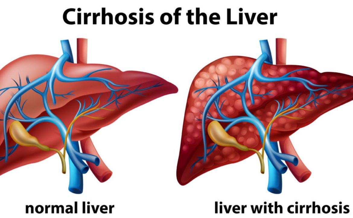
liver_cirrhosis
August 11, 2023 by Londonsono 0 Comments

Understanding Liver Cirrhosis: Causes, Symptoms, and Management

At our ultrasound clinic in London, we recognize the importance of educating our community about various liver conditions. Liver cirrhosis is a serious health concern that requires awareness, understanding, and timely management. In this article, we will delve into the causes, symptoms, and effective strategies for managing liver cirrhosis.

What is Liver Cirrhosis?

Liver cirrhosis is a progressive and irreversible condition in which healthy liver tissue is replaced by scar tissue. This scarring affects the liver’s normal structure and function, impairing its ability to perform vital tasks such as detoxification, protein synthesis, and bile production. If left untreated, cirrhosis can lead to severe complications, including liver failure.

Causes of Liver Cirrhosis:

Chronic Alcohol Abuse: Prolonged and excessive alcohol consumption is a leading cause of cirrhosis. Alcohol can directly damage liver cells and promote the accumulation of scar tissue.


Chronic Viral Hepatitis: Chronic infection with hepatitis B or C viruses can lead to ongoing inflammation and scarring of the liver, ultimately resulting in cirrhosis.

Non-Alcoholic Fatty Liver Disease (NAFLD): NAFLD, often associated with obesity and metabolic syndrome, involves the accumulation of fat in the liver. Over time, this can progress to cirrhosis.

Autoimmune Hepatitis: In this condition, the body’s immune system mistakenly attacks liver cells, causing inflammation and potential cirrhosis.

Genetic Disorders: Certain genetic conditions, such as hemochromatosis (excess iron accumulation) and Wilson’s disease (copper buildup), can contribute to cirrhosis.

Biliary Diseases: Conditions that affect the bile ducts, such as primary biliary cirrhosis and primary sclerosing cholangitis, can lead to cirrhosis.

Symptoms of Liver Cirrhosis:

In its early stages, cirrhosis may not cause noticeable symptoms. As the disease progresses, the following signs and symptoms may emerge:


Fatigue and weakness.

Loss of appetite.

Unexplained weight loss.

Nausea and vomiting.

Abdominal pain and discomfort.

Jaundice (yellowing of the skin and eyes).

Swelling in the legs and abdomen.

Itchy skin.

Importance of Early Detection:

Early detection and intervention are crucial for managing cirrhosis and preventing further liver damage. As a reputable ultrasound clinic in London, we offer advanced imaging techniques that aid in the early diagnosis and monitoring of liver health.

Diagnostic Tools:

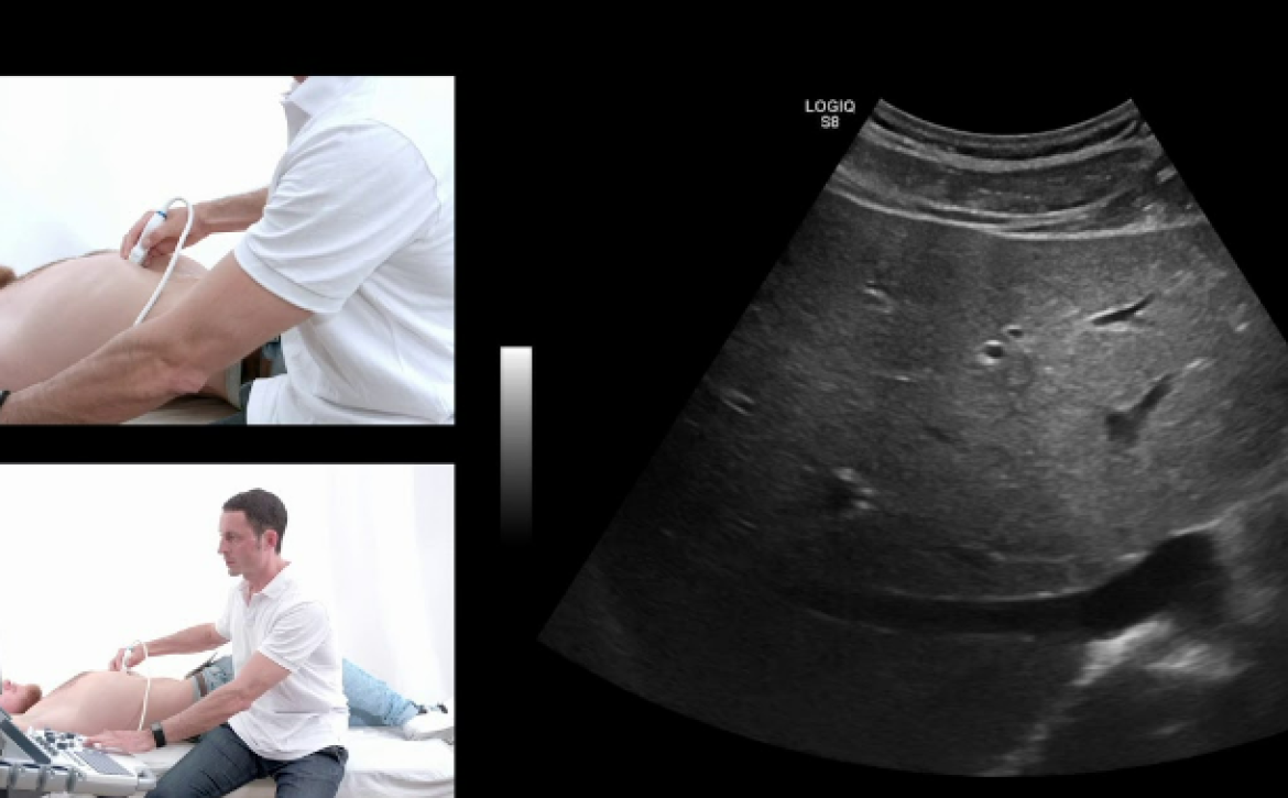
Ultrasound: Our high-resolution ultrasound imaging can detect changes in the liver’s structure, evaluate blood flow, and identify signs of cirrhosis.

FibroScan®: This non-invasive test measures liver stiffness, providing information about the degree of fibrosis (scarring) present in the liver.

Blood Tests: Specific blood markers can indicate liver function and the severity of cirrhosis.

Management and Treatment:

While cirrhosis is not reversible, effective management can slow its progression and alleviate symptoms:

Lifestyle Changes: If alcohol-related, abstaining from alcohol is crucial. Adopting a healthy diet, exercising regularly, and maintaining a healthy weight can improve liver health.

Medications: Depending on the underlying cause of cirrhosis, medications may be prescribed to manage complications, reduce inflammation, or address specific symptoms.

Regular Monitoring: Routine medical check-ups and follow-up appointments are essential to monitor the progression of cirrhosis and address any emerging issues.

Advanced Interventions: In some cases, advanced treatments such as liver transplantation may be considered for eligible patients with severe cirrhosis.

Prevention:

  • Preventing cirrhosis involves addressing its underlying causes and making healthy lifestyle choices:
  • Limit alcohol consumption or abstain from it entirely.
  • Get vaccinated against hepatitis B.
  • Practice safe sex to reduce the risk of hepatitis transmission.
  • Maintain a healthy weight and manage conditions like diabetes and high cholesterol.
  • Avoid sharing needles or other drug paraphernalia.

Conclusion:

Liver cirrhosis is a serious condition that demands attention, understanding, and early management. As a dedicated ultrasound clinic in London, we are committed to supporting our community’s liver health through advanced imaging techniques and comprehensive care. If you suspect you may be at risk for cirrhosis or are experiencing symptoms, don’t hesitate to contact our clinic for an evaluation. By raising awareness and taking proactive steps to address risk factors, we can collectively work towards preventing and effectively managing cirrhosis. Your liver health is our priority, and we are here to provide the support you

August 11, 2023 by Londonsono 0 Comments

Understanding Liver Cancer: Causes, Types, and Early Detection

Diagnosing Liver Cancer Symptoms

As a leading ultrasound clinic in London, we understand the importance of raising awareness about liver cancer, a condition that can significantly impact lives. Liver cancer, also known as hepatocellular carcinoma (HCC), is a serious disease that requires knowledge about its causes, types, and the crucial role of early detection for effective management and treatment.

Causes of Liver Cancer

Liver cancer often develops as a result of underlying liver disease, with certain risk factors increasing the likelihood of its occurrence:

Chronic Hepatitis Infections: Chronic infection with hepatitis B or C viruses significantly increases the risk of developing liver cancer. These infections can lead to inflammation and scarring of the liver, creating an environment conducive to cancerous growth.

Cirrhosis: Long-term liver damage and cirrhosis, whether due to alcohol abuse, fatty liver disease, or other causes, can elevate the risk of liver cancer.

Excessive Alcohol Consumption: Heavy and prolonged alcohol consumption can lead to cirrhosis, which in turn increases the risk of liver cancer.

Aflatoxin Exposure: Consumption of foods contaminated with aflatoxins, naturally occurring toxins produced by certain molds, can contribute to the development of liver cancer.

Non-Alcoholic Fatty Liver Disease (NAFLD): NAFLD, a condition characterized by excess fat in the liver, is also associated with an increased risk of liver cancer.

Types of Liver Cancer

Hepatocellular Carcinoma (HCC): This is the most common type of liver cancer and originates from the main type of liver cell (hepatocytes). It is often associated with chronic liver disease, cirrhosis, and hepatitis B or C infections.

Cholangiocarcinoma: This type of liver cancer develops in the bile ducts within the liver. It can obstruct bile flow and lead to jaundice and other symptoms.

Liver Cancer Symptoms of

Early-stage liver cancer may not cause noticeable symptoms. As the disease progresses, the following symptoms may occur:

Importance of Early Detection

Early detection of liver cancer is crucial for effective treatment and improved outcomes. As a trusted ultrasound clinic in London, we offer advanced imaging techniques that aid in the early diagnosis and monitoring of liver conditions, including cancer.

Diagnostic Tools:

Ultrasound: Ultrasound imaging can help detect changes in the liver’s structure and identify any suspicious growths or abnormalities.

CT Scan: A CT scan provides detailed cross-sectional images of the liver, helping to visualize tumors and their characteristics.

MRI: Magnetic resonance imaging (MRI) can provide high-resolution images that assist in evaluating the extent and location of liver tumors.

Liver Biopsy: In some cases, a small sample of liver tissue (biopsy) may be taken for analysis to confirm the presence of cancer.


Treatment Options:

Treatment for liver cancer depends on factors such as the type and stage of the cancer, as well as the patient’s overall health. Treatment options may include:


Surgery: Surgical removal of the tumor or even a liver transplant may be considered for eligible patients.

Ablation Therapy: Techniques such as radiofrequency ablation and microwave ablation use heat to destroy cancerous tissue.

Chemoembolization: This involves delivering chemotherapy directly to the tumor through the blood vessels that supply it.

Targeted Therapy: Targeted drugs are designed to inhibit specific molecules involved in cancer growth.

Immunotherapy: Immunotherapy helps the body’s immune system recognize and attack cancer cells.

Prevention:

While some risk factors for liver cancer are beyond our control, there are steps you can take to reduce your risk:

Get Vaccinated: If you’re at risk of hepatitis B, getting vaccinated can significantly lower your chances of developing liver cancer.

Practice Safe Sex: Reduce your risk of hepatitis B and C by practicing safe sex and using barrier methods.

Limit Alcohol Consumption: Moderating alcohol consumption can help protect your liver from damage.

Maintain a Healthy Weight: Obesity and fatty liver disease increase the risk of liver cancer. Maintaining a healthy weight through a balanced diet and regular exercise is essential.

Regular Check-ups: Regular medical check-ups and screenings are important, especially if you have risk factors for liver cancer.

Liver Fibroscan 

(Shearwave Elastography)

London Private Ultrasound Liver Cancer Clinics

Liver cancer is a serious condition that requires awareness, early detection, and timely intervention. As a dedicated ultrasound clinic in London, we are committed to assisting in the early diagnosis and monitoring of liver health. Our advanced imaging techniques, including ultrasound, CT scans, and MRI, play a vital role in detecting liver abnormalities, including cancer.


If you suspect any liver-related symptoms or have risk factors for liver cancer, don’t hesitate to reach out to our clinic for a comprehensive evaluation. Your liver health is our priority, and we are here to support you on your journey to optimal well-being

August 11, 2023 by Londonsono 0 Comments

Hepatitis B and C: Causes, Symptoms, and Importance of Early Detection


Hepatitis B and C are two types of viral infections that primarily affect the liver. They are a significant global health concern, with millions of people affected worldwide. As a leading ultrasound clinic in London, we understand the importance of raising awareness about these infections, their causes, symptoms, and the crucial role of early detection and management.

Hepatitis B:

Hepatitis B is caused by the hepatitis B virus (HBV) and is transmitted through contact with infected blood, semen, vaginal fluids, or other body fluids. It can be passed from an infected mother to her baby during childbirth, through sexual contact with an infected person, or by sharing needles and other drug-injection equipment. Here’s what you need to know about hepatitis B:

Causes of Hepatitis B

The cause of Hepatitis B is a blood bourne virus and can be spread via transmission through the blood or bodily fluids.

Unprotected sexual contact with an infected person.

Sharing needles or syringes with an infected person.

From mother to child during childbirth.

Accidental needlesticks or other sharp instrument injuries.

Symptoms:


  • Jaundice (yellowing of the skin and eyes).
  • Fatigue.
  • Abdominal pain.
  • Nausea and vomiting.
  • Dark urine and pale stools.
  • Joint pain.
  • Loss of appetite.

Hepatitis C

Hepatitis C is caused by the hepatitis C virus (HCV) and is primarily transmitted through contact with infected blood. It is a major cause of chronic liver disease, leading to cirrhosis and liver cancer. Understanding hepatitis C is crucial for preventing its spread and managing its impact:

Causes of Hepatitis C

The causes of hepatitis C are limited as the disease is blood bourne and therefore only  passed by blood.

Sharing needles or other drug paraphernalia.

Receiving a blood transfusion or organ transplant before widespread screening.

Needlestick injuries in healthcare settings.

Mother-to-child transmission during childbirth (less common).

Symptoms:

  • Jaundice (yellowing of the skin and eyes).

  • Fatigue.

  • Abdominal pain.

  • Loss of appetite.

  • Nausea and vomiting.

  • Dark urine and pale stools.

  • Joint pain.

Importance of Early Detection:

Both hepatitis B and C infections can be “silent” for many years, showing no noticeable symptoms. This makes regular screenings and early detection critically important, especially if you are at risk due to potential exposure. As a leading ultrasound clinic in London, we offer advanced imaging techniques that can assist in diagnosing and monitoring the progression of these infections.

Diagnostic Tools:

Ultrasound: London Private Ultrasound clinic high-resolution ultrasound imaging that can detect changes in the liver’s structure and identify any signs of liver damage or scarring (cirrhosis).

Blood Tests: Specific blood tests can identify markers of hepatitis B and C infections, helping us determine the presence and severity of the virus in your body.

FibroScan®: This non-invasive test measures liver stiffness, aiding in the assessment of liver fibrosis (scarring) caused by chronic hepatitis.

Prevention and Management:

Preventing hepatitis B and C involves adopting certain precautions:

  • Practice safe sex and use barrier methods.

  • Do not share needles, razors, or personal items that may have come into contact with blood.

  • Get vaccinated against hepatitis B.

  • Screen blood and organ donations.

  • Ensure proper sterilization of medical equipment.


Book Private Liver Fibroscan (shearwave Elastography)

Time is of the essence

Hepatitis B and C are serious viral infections that can lead to severe liver damage if left untreated. As a trusted ultrasound clinic in London, we emphasize the importance of awareness, early detection, and regular screenings for those at risk. Our advanced imaging techniques, including ultrasound and FibroScan®, play a crucial role in diagnosing, monitoring, and managing these infections.


Remember, early detection and proper medical guidance can lead to better outcomes and a healthier liver. If you suspect you may be at risk or need to schedule a screening, don’t hesitate to contact us. Your liver health is our priority and we can often provide same day appointments – available to book online.

August 11, 2023 by Londonsono 0 Comments

Understanding Fatty Liver: Causes, Symptoms, and Management

Fatty liver, medically known as hepatic steatosis, is a common condition characterized by the accumulation of excess fat within the liver cells. This condition can have various causes and can range from mild to more severe forms. Fatty liver is becoming increasingly prevalent worldwide, and it’s important to understand its causes, symptoms, and management strategies.

Causes of Fatty Liver:

Non-Alcoholic Fatty Liver Disease (NAFLD): The most common cause of fatty liver is non-alcoholic fatty liver disease, which is often associated with obesity, insulin resistance, and metabolic syndrome. These conditions contribute to the accumulation of fat within the liver cells.

Alcoholic Fatty Liver Disease: Excessive alcohol consumption over an extended period can lead to alcoholic fatty liver disease. Alcohol is processed in the liver, and heavy drinking can overwhelm the liver’s ability to metabolize fat.

Medications and Toxins: Certain medications, such as corticosteroids and tamoxifen, as well as exposure to toxins, can lead to fatty liver.

Rapid Weight Loss: Sudden and significant weight loss, especially in cases of obesity, can cause the liver to release fat into the bloodstream, leading to fatty liver.

Symptoms of Fatty Liver:

In its early stages, fatty liver may not cause noticeable symptoms. However, as the condition progresses, you may experience:

Fatigue

Unexplained weight loss

Pain or discomfort in the upper right abdomen

Enlarged liver

Elevated liver enzymes in blood tests

It’s important to note that these symptoms can be indicative of other conditions as well. Therefore, if you’re experiencing any of these symptoms, it’s crucial to consult a medical professional for proper evaluation.

If you are unsure of the symptoms you are experiencing we would recommend an abdominal ultrasound, this will allow the assessment of other possible causes of symptoms in the abdomen and liver such as diagnosing gallstones, tumours, cysts and also liver function.

Diagnosis and Risk Factors:

Fatty liver can be diagnosed through imaging studies such as ultrasound, CT scans, and MRI. These tests help visualize the amount of fat accumulated in the liver. Your doctor may also recommend blood tests to assess liver function and rule out other potential causes.

Certain factors increase the risk of developing fatty liver:

Obesity or overweight

Type 2 diabetes

High blood pressure

High cholesterol levels

Sedentary lifestyle

Poor diet

Rapid weight loss

Management and Prevention:

Managing and preventing fatty liver involves lifestyle modifications and, in some cases, medical intervention:

Healthy Diet: Adopting a balanced diet rich in fruits, vegetables, whole grains, lean proteins, and healthy fats can help prevent and manage fatty liver. Avoiding high-calorie, sugary, and processed foods is crucial.

Regular Exercise: Engaging in regular physical activity helps improve insulin sensitivity, aids in weight loss, and reduces fat accumulation in the liver.

Weight Management: Maintaining a healthy weight is essential for preventing and managing fatty liver. Gradual, sustainable weight loss can significantly improve liver health.

Limit Alcohol Consumption: If you have alcoholic fatty liver disease, reducing or eliminating alcohol consumption is crucial for recovery.

Medical Supervision: In some cases, medical intervention may be required. Your doctor may prescribe medications to manage underlying conditions, such as diabetes or high cholesterol.

How do you know if you have a fatty Liver?

Fatty liver is a prevalent condition that can have serious implications for your liver health. Understanding its causes, symptoms, and management strategies is essential for preventing its progression and ensuring optimal liver function. If you suspect you have fatty liver or are at risk, consult a medical professional for proper evaluation and guidance.

Remember, a healthy lifestyle, including a balanced diet, regular exercise, and avoiding excessive alcohol consumption, plays a key role in maintaining your liver’s well-being. Prioritising your liver health today can lead to a healthier tomorrow.

We offer a range of scans suitable for those wanting a diagnosis on fatty liver disease and the option below allows you to select your appointment date and time for a Liver Fibroscan additionally we offer a Liver and Pancreas screening blood test or a liver ultrasound option.

August 10, 2023 by Londonsono 0 Comments

Kidney Ultrasound Scans: Your window into kidney Health and Function

A kidney ultrasound scan is a valuable diagnostic procedure designed to assess the health of your kidneys, bladder (and prostate in men), and detect any potential abnormalities. Unlike CT scans that use radiation, this diagnostic investigation utilizes ultrasound waves for a safer imaging approach.

Booking a Kidney Ultrasound Scan

We understand the importance of convenience in scheduling your medical appointments. We offer a range of appointment options, including lunchtimes, evenings, and even weekends. To secure your preferred time and day, you can easily access our online booking diary. In case you encounter any challenges finding a suitable slot, our team is readily available through email or phone to assist you.

About Kidney Ultrasound

Ultrasound imaging, also known as sonography, involves using sound waves to create images of various parts of the body. It is a non-invasive and safe method, widely embraced for its efficacy in imaging organs, tissues, and even unborn babies. Ultrasound scans, performed by skilled sonographers, provide detailed images without the risks associated with radiation exposure.

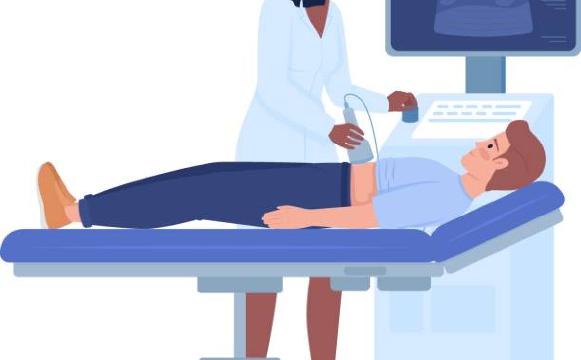
During a kidney ultrasound, a handheld probe is placed on the skin’s surface, emitting sound waves that penetrate the tissues. These waves bounce back to the transducer, and the resulting echoes are analyzed by a computer to create visual images on a screen. This process is akin to the principles of sonar technology used by the navy. Ultrasound is commonly used to visualize solid organs such as the liver, kidneys, uterus, and muscles, as well as to assess blood vessels and monitor fetal development during pregnancy.

Limitations of Renal Ultrasound Imaging

While renal ultrasound is a powerful diagnostic tool, it does have limitations. Ultrasound waves do not travel well through bowel or gas, which can hinder the evaluation of abdominal organs like the stomach and intestines. In addition, obtaining clear images from larger patients can be challenging due to the longer distance sound waves need to travel, resulting in weaker echoes.

Benefits and Risks of Kidney Ultrasound Examination

The benefits of undergoing a kidney ultrasound are numerous. It is a non-invasive procedure that is painless and safe for individuals of all ages, including children and expectant mothers. Ultrasound imaging is widely accessible, easy to use, and typically more affordable than other imaging modalities. Importantly, it doesn’t involve ionizing radiation, making it a safer choice.

There are virtually no risks associated with kidney ultrasound examinations. Unlike some medical procedures, ultrasound scans pose no harmful effects on humans.

Waiting Time and Access for Urinary Tract Ultrasound

The waiting time for an ultrasound scan appointment in NHS hospitals can vary, typically ranging from around six weeks. However, for those seeking more immediate attention and expedient access, our private scan clinic offers the possibility of same-day or next-day appointments for kidney ultrasound scans.

Understanding Kidney Ultrasound

Sound, which travels in waves, is the basis for ultrasound imaging. Ultrasound examinations involve creating images from the sound waves’ return as they encounter resistance within the body. A handheld sensor, resembling a microphone, is used to direct and capture these sound waves as they bounce back from various structures. The resulting images are examined by doctors and radiographers to diagnose conditions and guide medical procedures, including needle injections.

Detecting Kidney Stones and Cancer

Kidney ultrasound scans can effectively detect kidney stones and certain forms of cancer. While large stones are clearly visible and create distinctive shadows, smaller stones within the bladder can be challenging to visualize. Therefore, it’s crucial to have experienced sonographers conduct the scan to ensure accurate results. Similarly, kidney stones are visible on ultrasound scans, and their presence can be identified by the swelling of the renal pelvis caused by urine backflow.

Interpreting and Obtaining Results

The interpretation of kidney scan results is conducted by skilled healthcare professionals known as sonographers or ultrasonographers. These specialists perform the ultrasound scan, providing a diagnostic report of their findings. This report can then be shared with your doctor for further evaluation and care continuity. Throughout and after the examination, our sonographers are available to discuss results and address any inquiries.


Why Choose Us for Private Kidney Ultrasound


Our clinic, situated in the heart of London on Harley Street, offers a range of benefits for those seeking a kidney ultrasound. We provide same-day appointments within a clean and caring environment, ensuring your comfort and convenience. Our team of advanced consultant practitioners possesses extensive experience in ultrasound scanning. They are fully qualified, registered with respected healthcare organizations, and consistently updated with the latest technological trends and ultrasound guidelines. Your health and well-being are our priority, and our track record of satisfied clients and five-star reviews underscores our commitment to quality.


Our ultrasound clinic specializes in providing diagnostic private ultrasound scans, ensuring swift diagnoses and essential reassurance about your health. Located conveniently in central London, our commitment to affordability ensures that our services remain accessible without compromising quality. We believe that healthcare should be attainable for everyone, and our transparent pricing model reflects this principle. Our extensive client base, including GPs, consultants, radiologists, and midwives, speaks to the excellence of our services and the diagnostic accuracy we provide.


If you’re seeking a private kidney ultrasound scan, look no further. London Private Ultrasound Diagnostics offers a comprehensive range of ultrasound scans at the outset of Harley Street. With same-day and flexible out-of-hours appointments available, we cater to your busy schedule without compromising on the quality of care.

August 10, 2023 by Londonsono 0 Comments

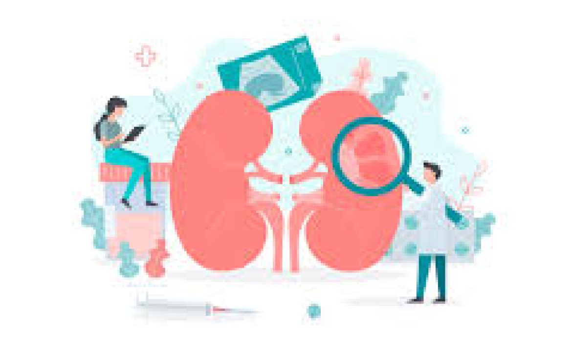
Kidney

At our ultrasound clinic in London, we offer a comprehensive Kidney Cancer Package to support the early detection and management of kidney cancer. Our dedicated team of professionals utilizes advanced ultrasound technology to provide accurate assessments. Here’s how we can assist you:

Blood and urine tests: Our clinic perform specialized blood and urine tests to detect any abnormalities that may indicate kidney cancer. These tests help assess levels of red blood cells, white blood cells, and various enzymes that can be indicative of the disease.

Kidney and Urinary Tract ultrasound: Our advanced ultrasound scans specifically focus on the kidneys and urinary tract. This non-invasive procedure allows us to detect the presence of tumors or nodules, determine their size, location, and assess whether they have spread to other parts of the body.

GP Consultation: We offer consultations with experienced general practitioners who specialize in kidney health. They will assess your symptoms, review your test results, and provide expert guidance and recommendations tailored to your specific needs.

Early detection and treatment are crucial for improving outcomes in kidney cancer. Our Kidney Cancer Package combines state-of-the-art ultrasound technology with expert medical care to ensure the best possible diagnosis and management.

If you have any concerns about kidney health or if you are at risk of kidney cancer, don’t hesitate to contact us and schedule an appointment. Your well-being is our priority, and we are here to support you every step of the way.