What Is the Difference Between Pelvic Scan and Abdominal Scan?
Medical imaging is critical in detecting and assessing a variety of disorders affecting the abdominal and pelvic areas. The abdominal and pelvic scans are two regularly used scans in this area. While both scans concentrate on different parts of the body, they serve separate functions and give significant information to healthcare practitioners. In this blog article, we’ll look at the distinctions between a pelvic scan and an abdominal scan in order to better comprehend their functions in medical imaging.
Scan of the Abdomen
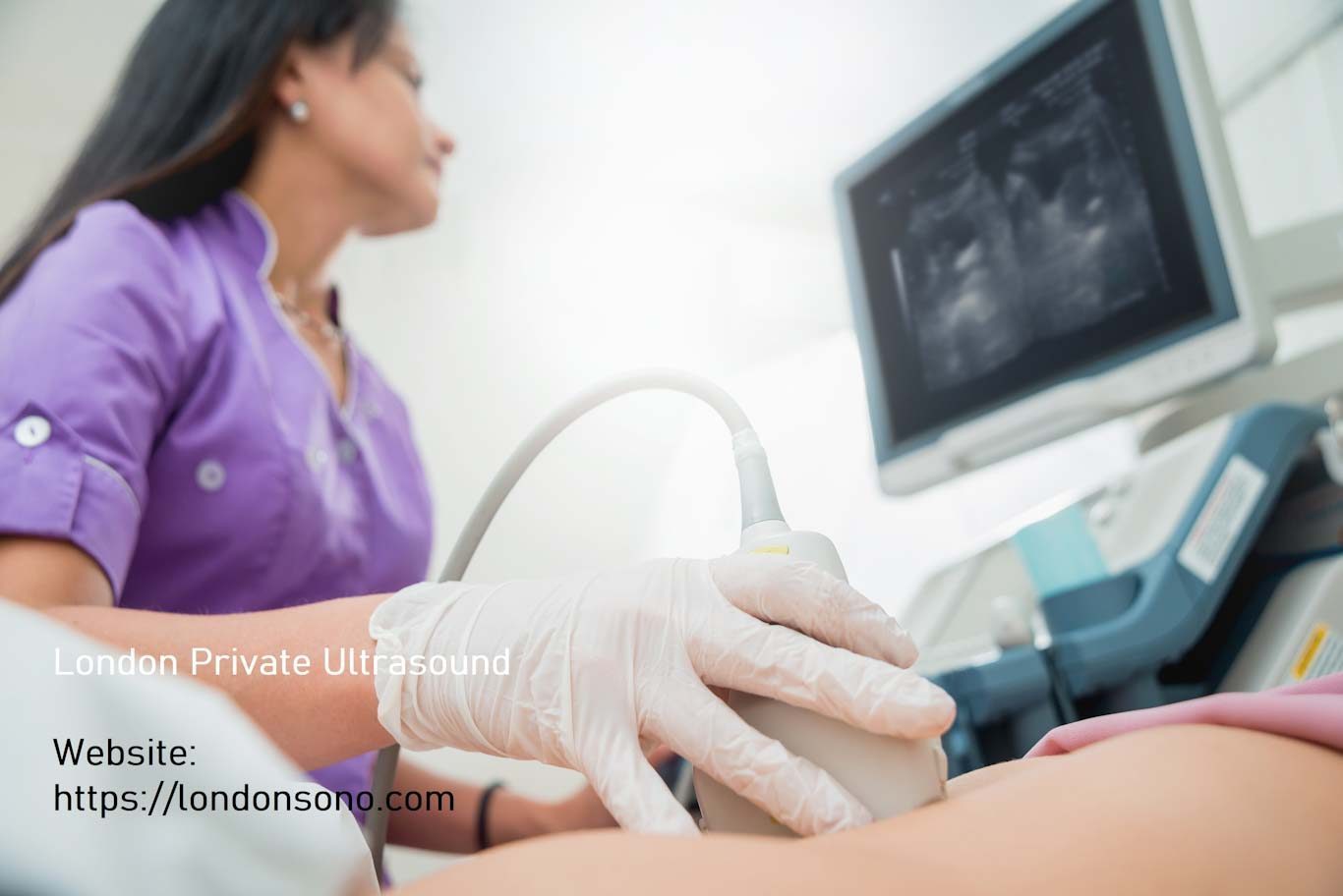
An abdominal scan, often known as an abdominal ultrasound, is a non-invasive imaging treatment that takes comprehensive pictures of the organs and structures in the belly. It uses high-frequency sound waves to generate real-time visuals on a display. A portable instrument called a transducer is softly pushed across the skin of the abdomen during an abdominal scan, producing sound waves and recording the returning echoes to make the pictures.
An abdominal scan’s primary goal is to assess the organs located in the abdominal cavity, which include the liver, gallbladder, pancreas, spleen, kidneys, and bladder, among others. It reveals important details on the size, shape, texture, and anomalies of these organs. A scan of the abdomen can aid in the diagnosis of illnesses such as liver disease, gallstones, pancreatic abnormalities, kidney stones, and abdominal tumours. It may also measure blood flow in the main arteries of the belly.
Scan of the Pelvic Organs
A pelvic scan, also known as a pelvic ultrasound, on the other hand, checks the organs and tissues in the pelvic area. The uterus, ovaries, fallopian tubes (in females), prostate gland (in men), bladder, and surrounding tissues are all part of the pelvic region. A pelvic scan, like an abdominal scan, uses sound waves and a transducer to produce real-time pictures.
A pelvic scan is used to assess the reproductive organs in both males and females. It aids in the diagnosis of disorders such as fibroids, ovarian cysts, polycystic ovary syndrome (PCOS), endometriosis, and pelvic inflammatory disease in females. It is also used during pregnancy to monitor foetal growth and assess the placenta. In men, a pelvic scan can detect problems such as benign prostatic hyperplasia (enlargement) or tumours in the prostate gland.
Distinctions and overlaps
The abdominal scan focuses on the organs of the abdominal cavity, whereas the pelvic scan focuses on the reproductive organs and associated tissues in the pelvic area. There is, however, some overlap between the two images. Both scans, for example, might look for abnormalities in the bladder, such as stones or tumours. To obtain a full examination of the abdominal and pelvic areas, a healthcare expert may propose a combination of both scans, known as a transabdominal and transvaginal ultrasound.
Finally, an abdominal scan and a pelvic scan are two independent imaging techniques that give important information about various parts of the body. The abdominal scan examines the organs in the abdominal cavity, whereas the pelvic scan examines the reproductive organs and associated tissues in the pelvic area. Both scans are non-invasive, safe, and often used to identify a wide range of disorders. If you have particular symptoms or concerns about your abdomen or pelvis, it is critical to contact with a healthcare specialist who can prescribe the best scan for your unique requirements.


